1DOR

DIHYDROOROTATE DEHYDROGENASE A FROM LACTOCOCCUS LACTIS


Experimental Data Snapshot

  • Method: X-RAY DIFFRACTION
  • Resolution: 2.00 Å
  • R-Value Free: 0.212 
  • R-Value Work: 0.168 
  • R-Value Observed: 0.168 

wwPDB Validation   3D Report Full Report


Ligand Structure Quality Assessment 


This is version 1.3 of the entry. See complete history


Literature

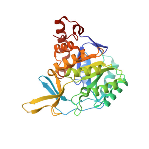
The crystal structure of the flavin containing enzyme dihydroorotate dehydrogenase A from Lactococcus lactis.

Rowland, P.Nielsen, F.S.Jensen, K.F.Larsen, S.

(1997) Structure 5: 239-252

  • DOI: https://doi.org/10.1016/s0969-2126(97)00182-2
  • Primary Citation of Related Structures:  
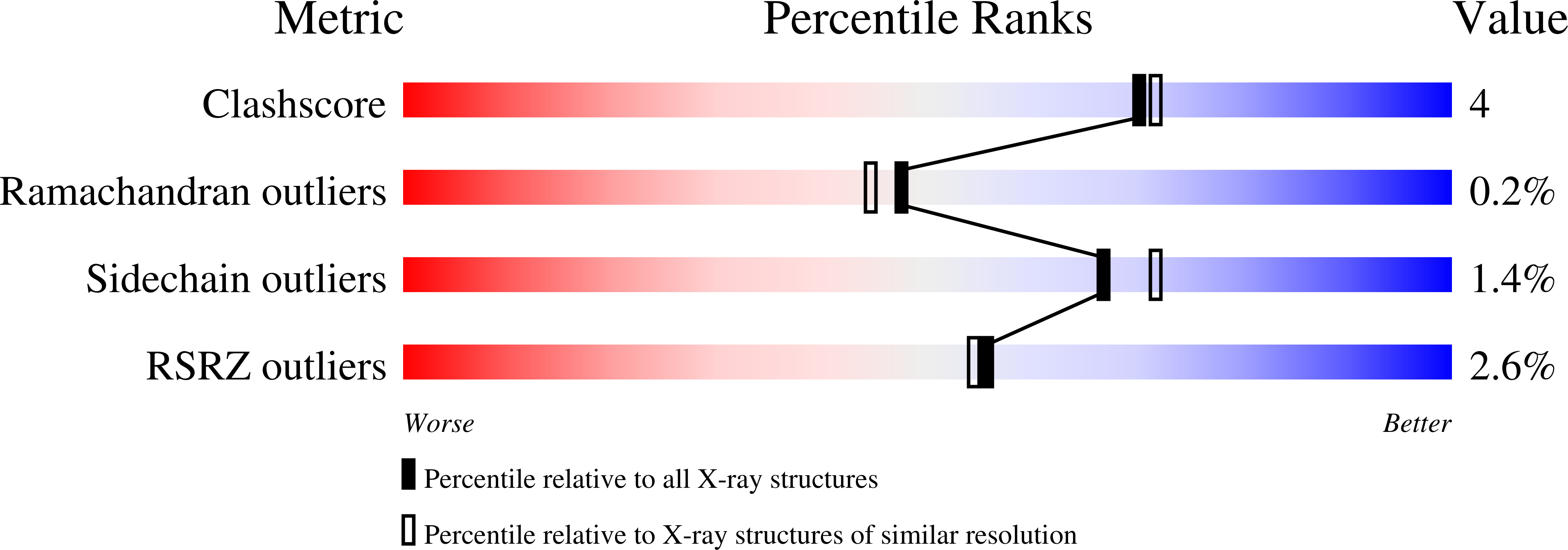
    1DOR

  • PubMed Abstract: 

    . Dihydroorotate dehydrogenase (DHOD) is a flavin mononucleotide containing enzyme, which catalyzes the oxidation of (S)-dihydroorotate to orotate, the fourth step in the de novo biosynthesis of pyrimidine nucleotides. Lactococcus lactis contains two genes encoding different functional DHODs whose sequences are only 30% identical. One of these enzymes, DHODA, is a highly efficient dimer, while the other, DHODB, shows optimal activity only in the presence of an iron-sulphur cluster containing protein with which it forms a complex tetramer. Sequence alignments have identified three different families among the DHODs: the two L. lactis enzymes belong to two of the families, whereas the enzyme from E. coli is a representative of the third. As no three-dimensional structures of DHODs are currently available, we set out to determine the crystal structure of DHODA from L. lactis. The differences between the two L. lactis enzymes make them particularly interesting for studying flavoprotein redox reactions and for identifying the differences between the enzyme families.


  • Organizational Affiliation

    Centre for Crystallographic Studies, Department of Chemistry University of Copenhagen, Universitetsparken 5, DK-2100 Copenhagen O, Denmark.


Macromolecules
Find similar proteins by:  (by identity cutoff)  |  3D Structure
Entity ID: 1
MoleculeChains Sequence LengthOrganismDetailsImage
DIHYDROOROTATE DEHYDROGENASE A
A, B
311Lactococcus lactisMutation(s): 0 
EC: 1.3.3.1
UniProt
Find proteins for A2RJT9 (Lactococcus lactis subsp. cremoris (strain MG1363))
Explore A2RJT9 
Go to UniProtKB:  A2RJT9
Entity Groups  
Sequence Clusters30% Identity50% Identity70% Identity90% Identity95% Identity100% Identity
UniProt GroupA2RJT9
Sequence Annotations
Expand
  • Reference Sequence
Small Molecules
Experimental Data & Validation

Experimental Data

  • Method: X-RAY DIFFRACTION
  • Resolution: 2.00 Å
  • R-Value Free: 0.212 
  • R-Value Work: 0.168 
  • R-Value Observed: 0.168 
  • Space Group: P 1 21 1
Unit Cell:
Length ( Å )Angle ( ˚ )
a = 54.19α = 90
b = 109.23β = 104.5
c = 67.17γ = 90
Software Package:
Software NamePurpose
X-PLORmodel building
X-PLORrefinement
DENZOdata reduction
SCALEPACKdata scaling
X-PLORphasing

Structure Validation

View Full Validation Report


Ligand Structure Quality Assessment 


Entry History 

Deposition Data

Revision History  (Full details and data files)

  • Version 1.0: 1997-04-21
    Type: Initial release
  • Version 1.1: 2008-03-24
    Changes: Version format compliance
  • Version 1.2: 2011-07-13
    Changes: Version format compliance
  • Version 1.3: 2024-02-07
    Changes: Data collection, Database references, Derived calculations, Other