1DLP

STRUCTURAL CHARACTERIZATION OF THE NATIVE FETUIN-BINDING PROTEIN SCILLA CAMPANULATA AGGLUTININ (SCAFET): A NOVEL TWO-DOMAIN LECTIN


Experimental Data Snapshot

  • Method: X-RAY DIFFRACTION
  • Resolution: 3.30 Å
  • R-Value Free: 0.293 
  • R-Value Work: 0.189 

wwPDB Validation   3D Report Full Report


This is version 1.3 of the entry. See complete history


Literature

Structural characterisation of the native fetuin-binding protein Scilla campanulata agglutinin: a novel two-domain lectin.

Wright, L.M.Reynolds, C.D.Rizkallah, P.J.Allen, A.K.Van Damme, E.J.Donovan, M.J.Peumans, W.J.

(2000) FEBS Lett 468: 19-22

  • DOI: https://doi.org/10.1016/s0014-5793(00)01109-1
  • Primary Citation of Related Structures:  
    1DLP

  • PubMed Abstract: 

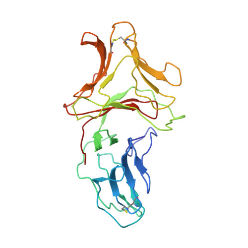
    The three-dimensional structure of a 244-residue, multivalent, fetuin-binding lectin, SCAfet, isolated from bluebell (Scilla campanulata) bulbs, has been solved at 3.3 A resolution by molecular replacement using the coordinates of the 119-residue, mannose-binding lectin, SCAman, also from bluebell bulbs. Unlike most monocot mannose-binding lectins, such as Galanthus nivalis agglutinin from snowdrop bulbs, which fold into a single domain, SCAfet contains two domains with approximately 55% sequence identity, joined by a linker peptide. Both domains are made up of a 12-stranded beta-prism II fold, with three putative carbohydrate-binding sites, one on each subdomain. SCAfet binds to the complex saccharides of various animal glycoproteins but not to simple sugars.


  • Organizational Affiliation

    School of Biomolecular Sciences, Max Perutz Building, Liverpool John Moores University, Liverpool, UK.


Macromolecules
Find similar proteins by:  (by identity cutoff)  |  3D Structure
Entity ID: 1
MoleculeChains Sequence LengthOrganismDetailsImage
LECTIN SCAFET PRECURSOR
A, B, C, D, E
A, B, C, D, E, F
236Hyacinthoides hispanicaMutation(s): 0 
UniProt
Find proteins for Q9ZP48 (Hyacinthoides hispanica)
Explore Q9ZP48 
Go to UniProtKB:  Q9ZP48
Entity Groups  
Sequence Clusters30% Identity50% Identity70% Identity90% Identity95% Identity100% Identity
UniProt GroupQ9ZP48
Sequence Annotations
Expand
  • Reference Sequence
Experimental Data & Validation

Experimental Data

  • Method: X-RAY DIFFRACTION
  • Resolution: 3.30 Å
  • R-Value Free: 0.293 
  • R-Value Work: 0.189 
  • Space Group: C 1 2 1
Unit Cell:
Length ( Å )Angle ( ˚ )
a = 277.94α = 90
b = 164.1β = 95.38
c = 53.6γ = 90
Software Package:
Software NamePurpose
MOSFLMdata reduction
ROTAVATAdata reduction
AMoREphasing
REFMACrefinement
CCP4data scaling
ROTAVATAdata scaling

Structure Validation

View Full Validation Report


Entry History 

Deposition Data

Revision History  (Full details and data files)

  • Version 1.0: 2000-02-10
    Type: Initial release
  • Version 1.1: 2008-04-27
    Changes: Version format compliance
  • Version 1.2: 2011-07-13
    Changes: Version format compliance
  • Version 1.3: 2017-10-04
    Changes: Advisory, Refinement description