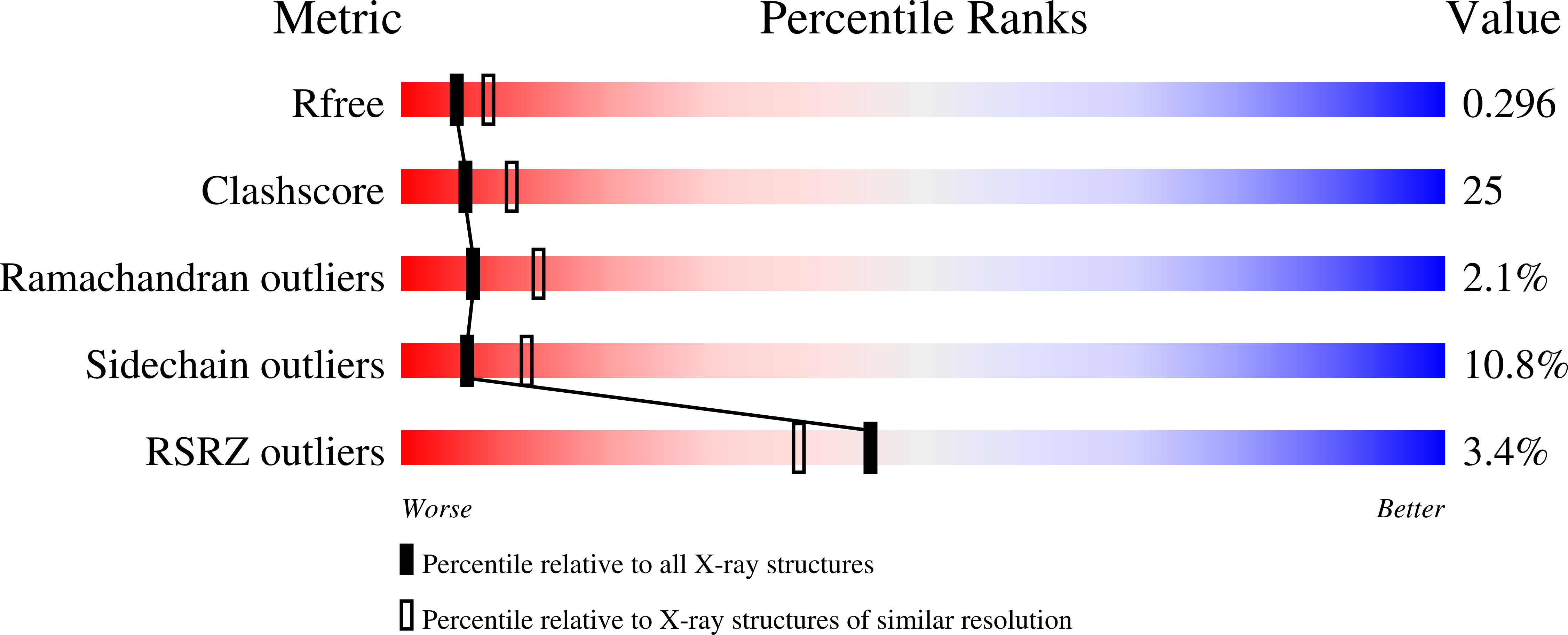
1DFQ

THE HC FRAGMENT OF TETANUS TOXIN COMPLEXED WITH SIALIC ACID


Experimental Data Snapshot

  • Method: X-RAY DIFFRACTION
  • Resolution: 2.60 Å
  • R-Value Free: 0.313 
  • R-Value Work: 0.200 
  • R-Value Observed: 0.200 

wwPDB Validation   3D Report Full Report


Ligand Structure Quality Assessment 


This is version 1.4 of the entry. See complete history


Literature

The structures of the H(C) fragment of tetanus toxin with carbohydrate subunit complexes provide insight into ganglioside binding.

Emsley, P.Fotinou, C.Black, I.Fairweather, N.F.Charles, I.G.Watts, C.Hewitt, E.Isaacs, N.W.

(2000) J Biol Chem 275: 8889-8894

  • DOI: https://doi.org/10.1074/jbc.275.12.8889
  • Primary Citation of Related Structures:  
    1D0H, 1DFQ, 1DIW, 1DLL

  • PubMed Abstract: 

    The entry of tetanus neurotoxin into neuronal cells proceeds through the initial binding of the toxin to gangliosides on the cell surface. The carboxyl-terminal fragment of the heavy chain of tetanus neurotoxin contains the ganglioside-binding site, which has not yet been fully characterized. The crystal structures of native H(C) and of H(C) soaked with carbohydrates reveal a number of binding sites and provide insight into the possible mode of ganglioside binding.


  • Organizational Affiliation

    Department of Chemistry, University of Glasgow, Glasgow G12 8QQ, United Kingdom.


Macromolecules
Find similar proteins by:  (by identity cutoff)  |  3D Structure
Entity ID: 1
MoleculeChains Sequence LengthOrganismDetailsImage
TETANUS TOXIN HC444Clostridium tetaniMutation(s): 0 
EC: 3.4.24.68
UniProt
Find proteins for P04958 (Clostridium tetani (strain Massachusetts / E88))
Explore P04958 
Go to UniProtKB:  P04958
Entity Groups  
Sequence Clusters30% Identity50% Identity70% Identity90% Identity95% Identity100% Identity
UniProt GroupP04958
Sequence Annotations
Expand
  • Reference Sequence
Small Molecules
Ligands 1 Unique
IDChains Name / Formula / InChI Key2D Diagram3D Interactions
SLB
Query on SLB

Download Ideal Coordinates CCD File 
B [auth A]N-acetyl-beta-neuraminic acid
C11 H19 N O9
SQVRNKJHWKZAKO-PFQGKNLYSA-N
Experimental Data & Validation

Experimental Data

  • Method: X-RAY DIFFRACTION
  • Resolution: 2.60 Å
  • R-Value Free: 0.313 
  • R-Value Work: 0.200 
  • R-Value Observed: 0.200 
  • Space Group: P 21 21 21
Unit Cell:
Length ( Å )Angle ( ˚ )
a = 66.86α = 90
b = 70.24β = 90
c = 122.52γ = 90
Software Package:
Software NamePurpose
AMoREphasing
REFMACrefinement
MOSFLMdata reduction
CCP4data scaling

Structure Validation

View Full Validation Report


Ligand Structure Quality Assessment 


Entry History 

Deposition Data

Revision History  (Full details and data files)

  • Version 1.0: 2000-03-24
    Type: Initial release
  • Version 1.1: 2008-04-27
    Changes: Version format compliance
  • Version 1.2: 2011-07-13
    Changes: Version format compliance
  • Version 1.3: 2020-07-29
    Type: Remediation
    Reason: Carbohydrate remediation
    Changes: Data collection, Derived calculations, Structure summary
  • Version 1.4: 2024-02-07
    Changes: Data collection, Database references, Structure summary