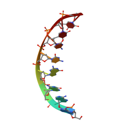
Structure refinement of the chromomycin dimer-DNA oligomer complex in solution.
Gao, X.L., Mirau, P., Patel, D.J.(1992) J Mol Biol 223: 259-279
- PubMed: 1731073
- DOI: https://doi.org/10.1016/0022-2836(92)90730-8
- Primary Citation of Related Structures:
1D83 - PubMed Abstract:
We have refined the initial docking model of the Mg(II)-co-ordinated chromomycin-d(A2G2C2T2) complex (2 drug equivalents per duplex) by a complete relaxation matrix analysis simulation of the two-dimensional nuclear Overhauser effect (NOESY) spectrum of the complex in 2H2O solution. This relaxation matrix refined structure of the complex exhibits the following characteristics. (1) We observe an unwound and elongated duplex that exhibits characteristics distinct from the A and B-DNA family of helices at the central (G-G-C-C).(G-G-C-C) chromomycin dimer binding and flanking sites. On the other hand sugar puckers, glycosidic torsion angles, displacement of the base-pairs from the helix axis and the minor groove width for this central tetranucleotide segment all fall within the A-family of helical parameters. (2) The chromomycin monomers are aligned in a head-to-tail orientation in the Mg(II)-co-ordinated dimer in the complex. The chromophores are aligned with a slight tilt relative to each other and make an angle of 75 degrees between their planes. The C-D-E trisaccharide segments from individual monomers adopt an extended conformation that projects in opposite directions in the dimer. The divalent metal cation is co-ordinated to the O(1) carbonyl and O(9) enolate atoms of the chromophores and aligns them such that the O(9)-Mg-O(9) angle is 170 degrees while all other O-Mg-O angles are in the 95(+/- 15)degrees range. (3) The sequence specificity of the chromomycin dimer for the widened and shallower (G3-G4-C5-C6).(G3-G4-C5-C6) minor groove binding site is associated with intermolecular hydrogen bonds formed between the OH group at C(8) of the chromophore and the minor groove NH2 group at position 2 and N(3) groups of G4 and between the O(1) oxygen of the E-sugar and the minor groove NH2 group at position 2 of G3 in the complex. (4) Additional intermolecular interactions are primarily van der Waals contacts between anomeric and adjacent CH2 protons on each sugar in the C-D-E trisaccharide segments of the chromomycin dimer and the minor groove surface of the DNA. These results provide insights into the induced conformational transitions required to generate a complementary match between the drug dimer and its DNA binding site on complex formation.
Organizational Affiliation:
Department of Biochemistry and Molecular Biophysics, College of Physicians and Surgeons, Columbia University, New York, N Y 10032.