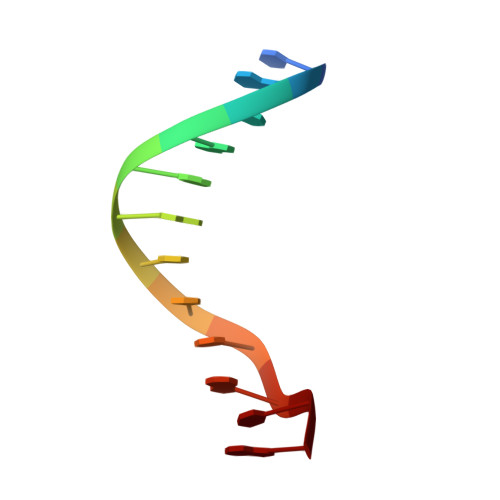
Crystal structure of a B-DNA dodecamer containing inosine, d(CGCIAATTCGCG), at 2.4 A resolution and its comparison with other B-DNA dodecamers.
Xuan, J.C., Weber, I.T.(1992) Nucleic Acids Res 20: 5457-5464
- PubMed: 1437563
- DOI: https://doi.org/10.1093/nar/20.20.5457
- Primary Citation of Related Structures:
1D77 - PubMed Abstract:
The crystal structure of the dodecamer, d(CGCIAATTCGCG), has been determined at 2.4 A resolution by molecular replacement, and refined to an R-factor of 0.174. The structure is isomorphous with that of the B-DNA dodecamer, d(CGCGAATTCGCG), in space group P2(1)2(1)2(1) with cell dimensions of a = 24.9, b = 40.4, and c = 66.4 A. The initial difference Fourier maps clearly indicated the presence of inosine instead of guanine. The structure was refined with 44 water molecules, and compared to the parent dodecamer. Overall the two structures are very similar, and the I:C forms Watson-Crick base pairs with similar hydrogen bond geometry to the G:C base pairs. The propeller twist angle is low for I4:C21 and relatively high for the I16:C9 base pair (-3.2 degrees compared to -23.0 degrees), and the buckle angles alter, probably due to differences in the contacts with symmetry related molecules in the crystal lattice. The central base pairs of d(CGCIAATTCGCG) show the large propeller twist angles, and the narrow minor groove that characterize A-tract DNA, although I:C base pairs cannot form the major groove bifurcated hydrogen bonds that are possible for A:T base pairs.
Organizational Affiliation:
Macromolecular Structure Laboratory, NCl-Frederick Cancer Research Facility, MD 21702.