1CVR

Crystal structure of the Arg specific cysteine proteinase gingipain R (RGPB)


Experimental Data Snapshot

  • Method: X-RAY DIFFRACTION
  • Resolution: 2.00 Å
  • R-Value Free: 0.207 
  • R-Value Work: 0.163 

wwPDB Validation   3D Report Full Report


Ligand Structure Quality Assessment 


This is version 1.4 of the entry. See complete history


Literature

Crystal structure of gingipain R: an Arg-specific bacterial cysteine proteinase with a caspase-like fold.

Eichinger, A.Beisel, H.G.Jacob, U.Huber, R.Medrano, F.J.Banbula, A.Potempa, J.Travis, J.Bode, W.

(1999) EMBO J 18: 5453-5462

  • DOI: https://doi.org/10.1093/emboj/18.20.5453
  • Primary Citation of Related Structures:  
    1CVR

  • PubMed Abstract: 

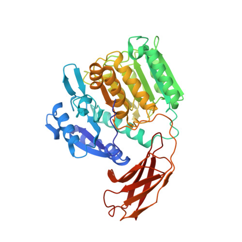
    Gingipains are cysteine proteinases acting as key virulence factors of the bacterium Porphyromonas gingivalis, the major pathogen in periodontal disease. The 1.5 and 2.0 A crystal structures of free and D-Phe-Phe-Arg-chloromethylketone-inhibited gingipain R reveal a 435-residue, single-polypeptide chain organized into a catalytic and an immunoglobulin-like domain. The catalytic domain is subdivided into two subdomains comprising four- and six-stranded beta-sheets sandwiched by alpha-helices. Each subdomain bears topological similarities to the p20-p10 heterodimer of caspase-1. The second subdomain harbours the Cys-His catalytic diad and a nearby Glu arranged around the S1 specificity pocket, which carries an Asp residue to enforce preference for Arg-P1 residues. This gingipain R structure is an excellent template for the rational design of drugs with a potential to cure and prevent periodontitis. Here we show the binding mode of an arginine-containing inhibitor in the active-site, thus identifying major interaction sites defining a suitable pharmacophor.


  • Organizational Affiliation

    Max-Planck-Institut für Biochemie, Abteilung Strukturforschung, D-82152 Martinsried, Germany.


Macromolecules
Find similar proteins by:  (by identity cutoff)  |  3D Structure
Entity ID: 1
MoleculeChains Sequence LengthOrganismDetailsImage
GINGIPAIN R435Porphyromonas gingivalisMutation(s): 0 
EC: 3.4.22.37
UniProt
Find proteins for P95493 (Porphyromonas gingivalis (strain ATCC BAA-308 / W83))
Explore P95493 
Go to UniProtKB:  P95493
Entity Groups  
Sequence Clusters30% Identity50% Identity70% Identity90% Identity95% Identity100% Identity
UniProt GroupP95493
Sequence Annotations
Expand
  • Reference Sequence
Small Molecules
Ligands 3 Unique
IDChains Name / Formula / InChI Key2D Diagram3D Interactions
H37
Query on H37

Download Ideal Coordinates CCD File 
B [auth A]D-phenylalanyl-N-[(3S)-6-carbamimidamido-1-chloro-2-oxohexan-3-yl]-L-phenylalaninamide
C25 H33 Cl N6 O3
PJFSUJMJVYGASC-HKBOAZHASA-N
ZN
Query on ZN

Download Ideal Coordinates CCD File 
H [auth A],
J [auth A]
ZINC ION
Zn
PTFCDOFLOPIGGS-UHFFFAOYSA-N
CA
Query on CA

Download Ideal Coordinates CCD File 
C [auth A]
D [auth A]
E [auth A]
F [auth A]
G [auth A]
C [auth A],
D [auth A],
E [auth A],
F [auth A],
G [auth A],
I [auth A]
CALCIUM ION
Ca
BHPQYMZQTOCNFJ-UHFFFAOYSA-N
Experimental Data & Validation

Experimental Data

  • Method: X-RAY DIFFRACTION
  • Resolution: 2.00 Å
  • R-Value Free: 0.207 
  • R-Value Work: 0.163 
  • Space Group: P 21 21 21
Unit Cell:
Length ( Å )Angle ( ˚ )
a = 51.93α = 90
b = 79.92β = 90
c = 99.82γ = 90
Software Package:
Software NamePurpose
MOLREPphasing
CNSrefinement
MOSFLMdata reduction
CCP4data scaling

Structure Validation

View Full Validation Report


Ligand Structure Quality Assessment 


Entry History 

Deposition Data

Revision History  (Full details and data files)

  • Version 1.0: 2000-03-01
    Type: Initial release
  • Version 1.1: 2008-04-27
    Changes: Version format compliance
  • Version 1.2: 2011-07-13
    Changes: Atomic model, Database references, Derived calculations, Non-polymer description, Structure summary, Version format compliance
  • Version 1.3: 2013-09-11
    Changes: Non-polymer description
  • Version 1.4: 2013-10-16
    Changes: Derived calculations, Non-polymer description