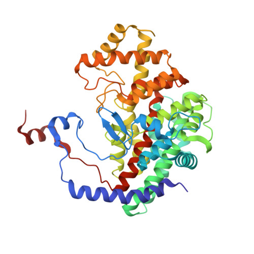
1CSH

A very short hydrogen bond provides only moderate stabilization of an enzyme: inhibitor complex of citrate synthase


Experimental Data Snapshot

  • Method: X-RAY DIFFRACTION
  • Resolution: 1.65 Å
  • R-Value Observed: 0.164 

wwPDB Validation   3D Report Full Report


This is version 2.0 of the entry. See complete history


Literature

A very short hydrogen bond provides only moderate stabilization of an enzyme-inhibitor complex of citrate synthase.

Usher, K.C.Remington, S.J.Martin, D.P.Drueckhammer, D.G.

(1994) Biochemistry 33: 7753-7759

  • DOI: https://doi.org/10.1021/bi00191a002
  • Primary Citation of Related Structures:  
    1CSH, 1CSI

  • PubMed Abstract: 

    Two extremely potent inhibitors of citrate synthase, carboxyl and primary amide analogues of acetyl coenzyme A, have been synthesized. The ternary complexes of these inhibitors with oxaloacetate and citrate synthase have been crystallized and their structures analyzed at 1.70- and 1.65-A resolution, respectively. The inhibitors have dissociation constants in the nanomolar range, with the carboxyl analogue binding more tightly (Ki = 1.6 nM at pH 6.0) than the amide analogue (28 nM), despite the unfavorable requirement for proton uptake by the former. The carboxyl group forms a shorter hydrogen bond with the catalytic Asp 375 (distance < 2.4 A) than does the amide group (distance approximately 2.5 A). Particularly with the carboxylate inhibitor, the very short hydrogen bond distances measured suggest a low barrier or short strong hydrogen bond. However, the binding constants differ by only a factor of 20 at pH 6.0, corresponding to an increase in binding energy for the carboxyl analogue on the enzyme of about 2 kcal/mol more than the amide analogue, much less than has been proposed for short strong hydrogen bonds based on gas phase measurements [> 20 kcal/mol (Gerlt & Gassman, 1993a,b)]. The inhibitor complexes support proposals that Asp 375 and His 274 work in concert to form an enolized form of acetyl-coenzyme A as the first step in the reaction.


  • Organizational Affiliation

    Institute of Molecular Biology, University of Oregon, Eugene 97403.


Macromolecules
Find similar proteins by:  (by identity cutoff)  |  3D Structure
Entity ID: 1
MoleculeChains Sequence LengthOrganismDetailsImage
CITRATE SYNTHASE435Gallus gallusMutation(s): 0 
EC: 4.1.3.7
UniProt
Find proteins for P23007 (Gallus gallus)
Explore P23007 
Go to UniProtKB:  P23007
Entity Groups  
Sequence Clusters30% Identity50% Identity70% Identity90% Identity95% Identity100% Identity
UniProt GroupP23007
Sequence Annotations
Expand
  • Reference Sequence
Small Molecules
Ligands 2 Unique
IDChains Name / Formula / InChI Key2D Diagram3D Interactions
AMX
Query on AMX

Download Ideal Coordinates CCD File 
C [auth A]AMIDOCARBOXYMETHYLDETHIA COENZYME *A
C23 H39 N8 O17 P3
RMWCNISNSABOOL-GRFIIANRSA-N
OAA
Query on OAA

Download Ideal Coordinates CCD File 
B [auth A]OXALOACETATE ION
C4 H3 O5
KHPXUQMNIQBQEV-UHFFFAOYSA-M
Binding Affinity Annotations 
IDSourceBinding Affinity
AMX Binding MOAD:  1CSH Ki: 28 (nM) from 1 assay(s)
PDBBind:  1CSH Ki: 28 (nM) from 1 assay(s)
Experimental Data & Validation

Experimental Data

  • Method: X-RAY DIFFRACTION
  • Resolution: 1.65 Å
  • R-Value Observed: 0.164 
  • Space Group: C 1 2 1
Unit Cell:
Length ( Å )Angle ( ˚ )
a = 104.4α = 90
b = 78.5β = 78.9
c = 58.5γ = 90
Software Package:
Software NamePurpose
TNTrefinement

Structure Validation

View Full Validation Report


Entry History 

Deposition Data

Revision History  (Full details and data files)

  • Version 1.0: 1994-04-30
    Type: Initial release
  • Version 1.1: 2008-03-03
    Changes: Version format compliance
  • Version 1.2: 2011-07-13
    Changes: Version format compliance
  • Version 1.3: 2017-11-29
    Changes: Derived calculations, Other
  • Version 2.0: 2024-02-07
    Changes: Atomic model, Data collection, Database references, Derived calculations