1CP8

NMR STRUCTURE OF DNA (5'-D(TTGGCCAA)2-3') COMPLEXED WITH NOVEL ANTITUMOR DRUG UCH9


Experimental Data Snapshot

  • Method: SOLUTION NMR
  • Conformers Submitted: 
  • Selection Criteria: LEAST RESTRAINT VIOLATION 

wwPDB Validation   3D Report Full Report


This is version 2.1 of the entry. See complete history


Literature

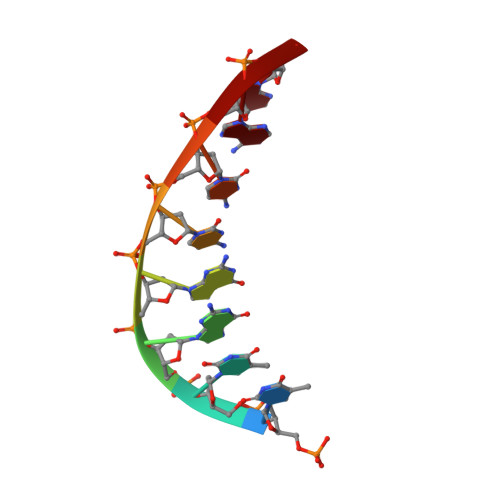
Solution structure of the novel antitumor drug UCH9 complexed with d(TTGGCCAA)2 as determined by NMR.

Katahira, R.Katahira, M.Yamashita, Y.Ogawa, H.Kyogoku, Y.Yoshida, M.

(1998) Nucleic Acids Res 26: 744-755

  • DOI: https://doi.org/10.1093/nar/26.3.744
  • Primary Citation of Related Structures:  
    1CP8

  • PubMed Abstract: 

    Aureolic acid group compounds, such as chromomycin A3(CHM) and mithramycin (MIT), are known as antitumor drugs. Recently we isolated a novel aureolic acid group antitumor drug, UCH9, from Streptomyces sp. The chemical structure of UCH9 is unique in that mono- (A ring) and tetrasaccharide (B-E rings) segments and a longer hydrophobic sidechain are attached to the chromophore, while di- and trisaccharide segments and a methyl group are attached to it in the cases of CHM and MIT. It has been shown by two-dimensional agarose gel electrophoresis that the three drugs cause DNA unwinding, UCH9 causing less than the others. A photo-CIDNP experiment has revealed that UCH9 binds to the minor groove of DNA. The structure of the UCH9-d(TTGGCCAA)2 complex has been determined by 1H NMR and simulated annealing calculations. The obtained structure indicates that UCH9 binds as a dimer to the minor groove of d(TTGGCCAA)2, like CHM and MIT, but that the structural change in DNA induced on binding of UCH9 is moderate in comparison with those on binding of the other two drugs. It turns out that the dimer structure of UCH9, stabilized presumably through a hydrophobic interaction involving the A, D and E rings and the hydrophobic sidechain is different from that of CHM and thus DNA can interact with UCH9 in the minor groove with a moderate structural change.


  • Organizational Affiliation

    Tokyo Research Laboratories, Kyowa Hakko Kogyo Co. Ltd, 3-6-6 Asahimachi, Machida-shi, Tokyo 194, Japan.


Macromolecules

Find similar nucleic acids by:  Sequence   |   3D Structure  

Entity ID: 1
MoleculeChains LengthOrganismImage
DNA (5'-D(P*TP*TP*GP*GP*CP*CP*AP*A)-3')
A, B
8N/A
Sequence Annotations
Expand
  • Reference Sequence
Oligosaccharides

Help

Entity ID: 2
MoleculeChains Length2D Diagram Glycosylation3D Interactions
beta-D-Olivopyranose-(1-3)-2,6-dideoxy-4-O-methyl-beta-D-glucopyranose-(1-3)-2,6-dideoxy-beta-D-galactopyranose-(1-3)-beta-D-Olivopyranose
C, D
4N/A
Glycosylation Resources
GlyTouCan:  G59463XK
GlyCosmos:  G59463XK
Small Molecules
Ligands 3 Unique
IDChains Name / Formula / InChI Key2D Diagram3D Interactions
DXA
Query on DXA

Download Ideal Coordinates CCD File 
F [auth A],
I [auth B]
1,2-HYDRO-1-OXY-3,4-HYDRO-3-(1-METHOXY-2-OXY-3,4-DIHYDROXYPENTYL)-8,9-DIHYDROXY-7-(SEC-BUTYL)-ANTHRACENE
C24 H30 O7
XGORHSPGULYMCV-HHYQQTLHSA-N
DDA
Query on DDA

Download Ideal Coordinates CCD File 
E [auth A],
G [auth B]
beta-D-Olivopyranose
C6 H12 O4
FDWRIIDFYSUTDP-KVTDHHQDSA-N
MG
Query on MG

Download Ideal Coordinates CCD File 
H [auth B]MAGNESIUM ION
Mg
JLVVSXFLKOJNIY-UHFFFAOYSA-N
Experimental Data & Validation

Experimental Data

  • Method: SOLUTION NMR
  • Conformers Submitted: 
  • Selection Criteria: LEAST RESTRAINT VIOLATION 

Structure Validation

View Full Validation Report


Entry History 

Deposition Data

Revision History  (Full details and data files)

  • Version 1.0: 1999-07-01
    Type: Initial release
  • Version 1.1: 2008-04-26
    Changes: Version format compliance
  • Version 1.2: 2011-07-13
    Changes: Version format compliance
  • Version 2.0: 2020-07-29
    Type: Remediation
    Reason: Carbohydrate remediation
    Changes: Atomic model, Data collection, Derived calculations, Structure summary
  • Version 2.1: 2023-12-27
    Changes: Data collection, Database references, Structure summary