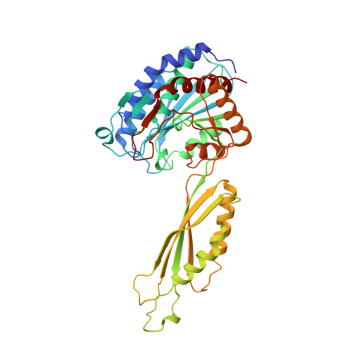
Crystal structure of carboxypeptidase G2, a bacterial enzyme with applications in cancer therapy.
Rowsell, S., Pauptit, R.A., Tucker, A.D., Melton, R.G., Blow, D.M., Brick, P.(1997) Structure 5: 337-347
- PubMed: 9083113
- DOI: https://doi.org/10.1016/s0969-2126(97)00191-3
- Primary Citation of Related Structures:
1CG2 - PubMed Abstract:
Carboxypeptidase G enzymes hydrolyze the C-terminal glutamate moiety from folic acid and its analogues, such as methotrexate. The enzyme studied here, carboxypeptidase G2 (CPG2), is a dimeric zinc-dependent exopeptidase produced by Pseudomonas sp. strain RS-16. CPG2 has applications in cancer therapy: following its administration as an immunoconjugate, in which CPG2 is linked to an antibody to a tumour-specific antigen, it can enzymatically convert subsequently administered inactive prodrugs to cytotoxic drugs selectively at the tumour site. CPG2 has no significant amino acid sequence homology with proteins of known structure. Hence, structure determination of CPG2 was undertaken to identify active-site residues, which may in turn provide ideas for protein and/or substrate modification with a view to improving its therapeutic usefulness. We have determined the crystal structure of CPG2 at 2.5 A resolution using multiple isomorphous replacement methods and non-crystallographic symmetry averaging. Each subunit of the molecular dimer consists of a larger catalytic domain containing two zinc ions at the active site, and a separate smaller domain that forms the dimer interface. The two active sites in the dimer are more than 60 A apart and are presumed to be independent; each contains a symmetric distribution of carboxylate and histidine ligands around two zinc ions which are 3.3 A apart. This distance is bridged by two shared zinc ligands, an aspartic acid residue and a hydroxyl ion. We find that the CPG2 catalytic domain has structural homology with other zinc-dependent exopeptidases, both those with a single zinc ion and those with a pair of zinc ions in the active site. The closest structural homology is with the aminopeptidase from Aeromonas proteolytica, where the similarity includes superposable zinc ligands but does not extend to the rest of the active-site residues, consistent with the different substrate specificities. The mechanism of peptide cleavage is likely to be very similar in these two enzymes and may involve the bridging hydroxyl ion ligand acting as a primary nucleophile.
Organizational Affiliation:
Blackett Laboratory, Imperial College London, SW7 2BZ, UK.