Phe161 and Arg166 variants of p-hydroxybenzoate hydroxylase. Implications for NADPH recognition and structural stability.
Eppink, M.H., Bunthol, C., Schreuder, H.A., van Berkel, W.J.(1999) FEBS Lett 443: 251-255
- PubMed: 10025942
- DOI: https://doi.org/10.1016/s0014-5793(98)01726-8
- Primary Citation of Related Structures:
1CC4, 1CC6 - PubMed Abstract:
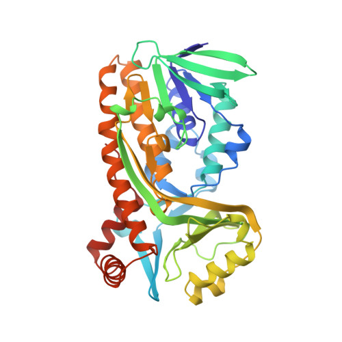
Phe161 and Arg166 of p-hydroxybenzoate hydroxylase from Pseudomonas fluorescens belong to a newly discovered sequence motif in flavoprotein hydroxylases with a putative dual function in FAD and NADPH binding [1]. To study their role in more detail, Phe161 and Arg166 were selectively changed by site-directed mutagenesis. F161A and F161G are catalytically competent enzymes having a rather poor affinity for NADPH. The catalytic properties of R166K are similar to those of the native enzyme. R166S and R166E show impaired NADPH binding and R166E has lost the ability to bind FAD. The crystal structure of substrate complexed F161A at 2.2 A is indistinguishable from the native enzyme, except for small changes at the site of mutation. The crystal structure of substrate complexed R166S at 2.0 A revealed that Arg166 is important for providing an intimate contact between the FAD binding domain and a long excursion of the substrate binding domain. It is proposed that this interaction is essential for structural stability and for the recognition of the pyrophosphate moiety of NADPH.
Organizational Affiliation:
Department of Biomolecular Sciences, Wageningen University Research Centre, The Netherlands.