1C53

S-CLASS CYTOCHROMES C HAVE A VARIETY OF FOLDING PATTERNS: STRUCTURE OF CYTOCHROME C-553 FROM DESULFOVIBRIO VULGARIS DETERMINED BY THE MULTI-WAVELENGTH ANOMALOUS DISPERSION METHOD


Experimental Data Snapshot

  • Method: X-RAY DIFFRACTION
  • Resolution: 1.80 Å
  • R-Value Observed: 0.194 

wwPDB Validation   3D Report Full Report


This is version 1.3 of the entry. See complete history


Literature

S-class cytochromes c have a variety of folding patterns: structure of cytochrome c-553 from Desulfovibrio vulgaris determined by the multi-wavelength anomalous dispersion method.

Nakagawa, A.Higuchi, Y.Yasuoka, N.Katsube, Y.Yagi, T.

(1990) J Biochem 108: 701-703

  • DOI: https://doi.org/10.1093/oxfordjournals.jbchem.a123267
  • Primary Citation of Related Structures:  
    1C53

  • PubMed Abstract: 

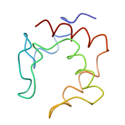
    The three-dimensional structure of cytochrome c-553 isolated from sulfate-reducing bacterium, Desulfovibrio vulgaris Miyazaki F strain, has been determined by the multi-wavelength anomalous dispersion technique with use of synchrotron radiation. The result shows that bacterial S-class cytochromes c have a variety of folding patterns. The relative location of two a-helices at amino- and carboxyl-terminals and the style of bonding to the heme group show "cytochrome c folding," but other regions of the structure are different from those of other cytochromes c previously reported. The results also give useful information about the location of sulfate-reducing bacterium on the phylogenetic tree of the bacterial cytochromes c superfamily.


  • Organizational Affiliation

    Photon Factory, National Laboratory for High Energy Physics, Hyogo.


Macromolecules
Find similar proteins by:  (by identity cutoff)  |  3D Structure
Entity ID: 1
MoleculeChains Sequence LengthOrganismDetailsImage
CYTOCHROME C55379Nitratidesulfovibrio vulgaris str. 'Miyazaki FMutation(s): 0 
UniProt
Find proteins for P00120 (Desulfovibrio vulgaris (strain DSM 19637 / Miyazaki F))
Explore P00120 
Go to UniProtKB:  P00120
Entity Groups  
Sequence Clusters30% Identity50% Identity70% Identity90% Identity95% Identity100% Identity
UniProt GroupP00120
Sequence Annotations
Expand
  • Reference Sequence
Small Molecules
Ligands 1 Unique
IDChains Name / Formula / InChI Key2D Diagram3D Interactions
HEM
Query on HEM

Download Ideal Coordinates CCD File 
B [auth A]PROTOPORPHYRIN IX CONTAINING FE
C34 H32 Fe N4 O4
KABFMIBPWCXCRK-RGGAHWMASA-L
Experimental Data & Validation

Experimental Data

  • Method: X-RAY DIFFRACTION
  • Resolution: 1.80 Å
  • R-Value Observed: 0.194 
  • Space Group: P 43 21 2
Unit Cell:
Length ( Å )Angle ( ˚ )
a = 42.7α = 90
b = 42.7β = 90
c = 103.4γ = 90
Software Package:
Software NamePurpose
PROLSQrefinement

Structure Validation

View Full Validation Report


Entry History 

Deposition Data

Revision History  (Full details and data files)

  • Version 1.0: 1993-10-31
    Type: Initial release
  • Version 1.1: 2008-03-24
    Changes: Version format compliance
  • Version 1.2: 2011-07-13
    Changes: Version format compliance
  • Version 1.3: 2024-02-07
    Changes: Data collection, Database references, Derived calculations, Other