1BHD

SECOND CALPONIN HOMOLOGY DOMAIN FROM UTROPHIN


Experimental Data Snapshot

  • Method: X-RAY DIFFRACTION
  • Resolution: 2.00 Å
  • R-Value Free: 0.257 
  • R-Value Work: 0.185 
  • R-Value Observed: 0.202 

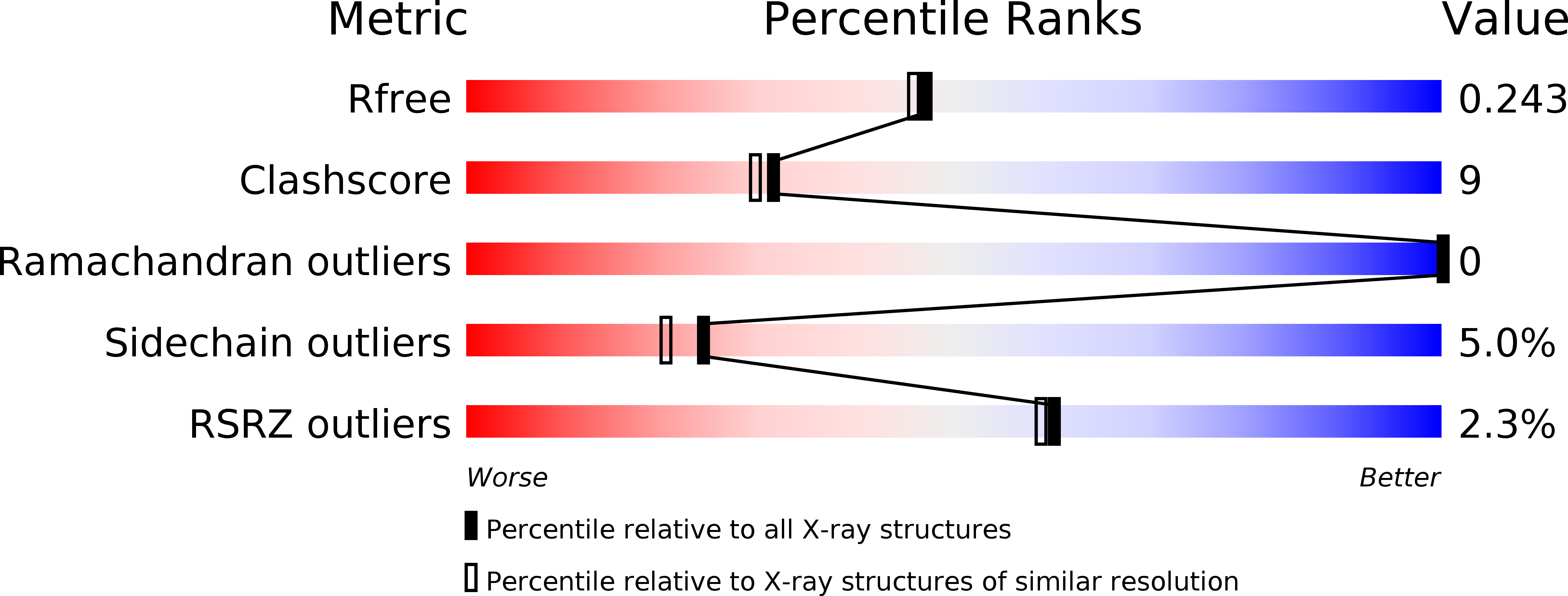
wwPDB Validation   3D Report Full Report


This is version 1.4 of the entry. See complete history


Literature

The 2.0 A structure of the second calponin homology domain from the actin-binding region of the dystrophin homologue utrophin.

Keep, N.H.Norwood, F.L.Moores, C.A.Winder, S.J.Kendrick-Jones, J.

(1999) J Mol Biol 285: 1257-1264

  • DOI: https://doi.org/10.1006/jmbi.1998.2406
  • Primary Citation of Related Structures:  
    1BHD

  • PubMed Abstract: 

    Utrophin is a close homologue of dystrophin, the protein defective in Duchenne muscular dystrophy. Like dystrophin, it is composed of three regions: an N-terminal region that binds actin filaments, a large central region with triple coiled-coil repeats, and a C-terminal region that interacts with components in the dystroglycan protein complex at the plasma membrane. The N-terminal actin-binding region consists of two calponin homology domains and is related to the actin-binding domains of a superfamily of proteins including alpha-actinin, spectrin and fimbrin. Here, we present the 2.0 A structure of the second calponin homology domain of utrophin solved by X-ray crystallography, and compare it to the other calponin homology domains previously determined from spectrin and fimbrin.


  • Organizational Affiliation

    MRC Laboratory of Molecular Biology, Hills Road, Cambridge, CB2 2QH, UK. n.keep@mail.cryst.bbk.ac.uk


Macromolecules
Find similar proteins by:  (by identity cutoff)  |  3D Structure
Entity ID: 1
MoleculeChains Sequence LengthOrganismDetailsImage
UTROPHIN
A, B
118Homo sapiensMutation(s): 0 
Gene Names: UTRNDMDL
UniProt & NIH Common Fund Data Resources
Find proteins for P46939 (Homo sapiens)
Explore P46939 
Go to UniProtKB:  P46939
PHAROS:  P46939
GTEx:  ENSG00000152818 
Entity Groups  
Sequence Clusters30% Identity50% Identity70% Identity90% Identity95% Identity100% Identity
UniProt GroupP46939
Sequence Annotations
Expand
  • Reference Sequence
Experimental Data & Validation

Experimental Data

  • Method: X-RAY DIFFRACTION
  • Resolution: 2.00 Å
  • R-Value Free: 0.257 
  • R-Value Work: 0.185 
  • R-Value Observed: 0.202 
  • Space Group: P 1 21 1
Unit Cell:
Length ( Å )Angle ( ˚ )
a = 63.92α = 90
b = 32.21β = 116.25
c = 65.36γ = 90
Software Package:
Software NamePurpose
AMoREphasing
REFMACrefinement
MOSFLMdata reduction
CCP4data scaling

Structure Validation

View Full Validation Report


Entry History 

Deposition Data

Revision History  (Full details and data files)

  • Version 1.0: 1999-01-13
    Type: Initial release
  • Version 1.1: 2008-03-24
    Changes: Version format compliance
  • Version 1.2: 2011-07-13
    Changes: Version format compliance
  • Version 1.3: 2018-05-30
    Changes: Data collection, Derived calculations, Experimental preparation
  • Version 1.4: 2023-08-02
    Changes: Database references, Refinement description