High resolution crystal structure of the A-DNA decamer d(CCCGGCCGGG). Novel intermolecular base-paired G*(G.C) triplets.
Ramakrishnan, B., Sundaralingam, M.(1993) J Mol Biol 231: 431-444
- PubMed: 8510155
- DOI: https://doi.org/10.1006/jmbi.1993.1292
- Primary Citation of Related Structures:
160D - PubMed Abstract:
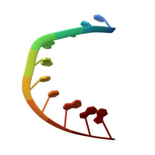
The DNA decamer d(CCCGGCCGGG) crystallizes in the orthorhombic space group P2(1)2(1)2(1) with a = 24.88, b = 44.60 and c = 46.97 A containing a duplex in the asymmetric unit. The structure was solved by molecular replacement and refined to an R factor of 18.5% using 6033 reflections at 1.65 A resolution. The decamer duplex adopts an A-DNA conformation. The abrupt dislocation of the duplex at the fourth base-pair G(4).C(17) by an abutting symmetry related molecule results in distortion of the backbone bonds of the fifth residue G(5), P-O(5')(alpha) and C(4')-C(5')(gamma), to the trans conformations from their favored gauche- and gauche+ conformations, respectively. In this close encounter the terminal G(10).C(11) base-pair of the symmetry related molecule hydrogen bonds to the G(4).C(17) base-pair forming a novel base-paired G(4)*(G10).C(11)) triplet, where G(4) is hydrogen bonded to both G(10) and C(11). To facilitate this hydrogen bonding the G(4).C(17) base-pair slides into the minor groove, causing a toll on the backbone conformation of the adjacent residue G(5). A similar triplet base-pairing interaction with somewhat weaker hydrogen bonds occurs at the pseudo dyad related C(7).G(14) base-pair with G(20) of another symmetry related duplex. This pseudo triplet interaction (C(7).G(14))*G(20), does not perturb the backgone alpha and gamma torsions of G(15). Both the novel base triplets are non-planar. The abrupt dislocation/bend at the G(4).C(17) base-pair jolts the global helical base-pair parameters, inclination, tilt, roll, tip, etc. quite markedly. Therefore a better description of the helix parameters is obtained by splitting the duplex and calculating the local helix axis for the top half consisting of the first three base-pairs, and the lower half consisting of the last six base-pairs, omitting the fourth base-pair. The two half duplexes are bent by only 10 degrees. This structure further demonstrates that crystal packing interactions, which can also be governed by base sequence, play a dominant role in determining DNA conformation.
Organizational Affiliation:
Department of Chemistry, Ohio State University, Columbus 43210-1002.