


Crystal structure of Langat virus helicase
Chen, C., Xue, R.L.To be published.
Experimental Data Snapshot
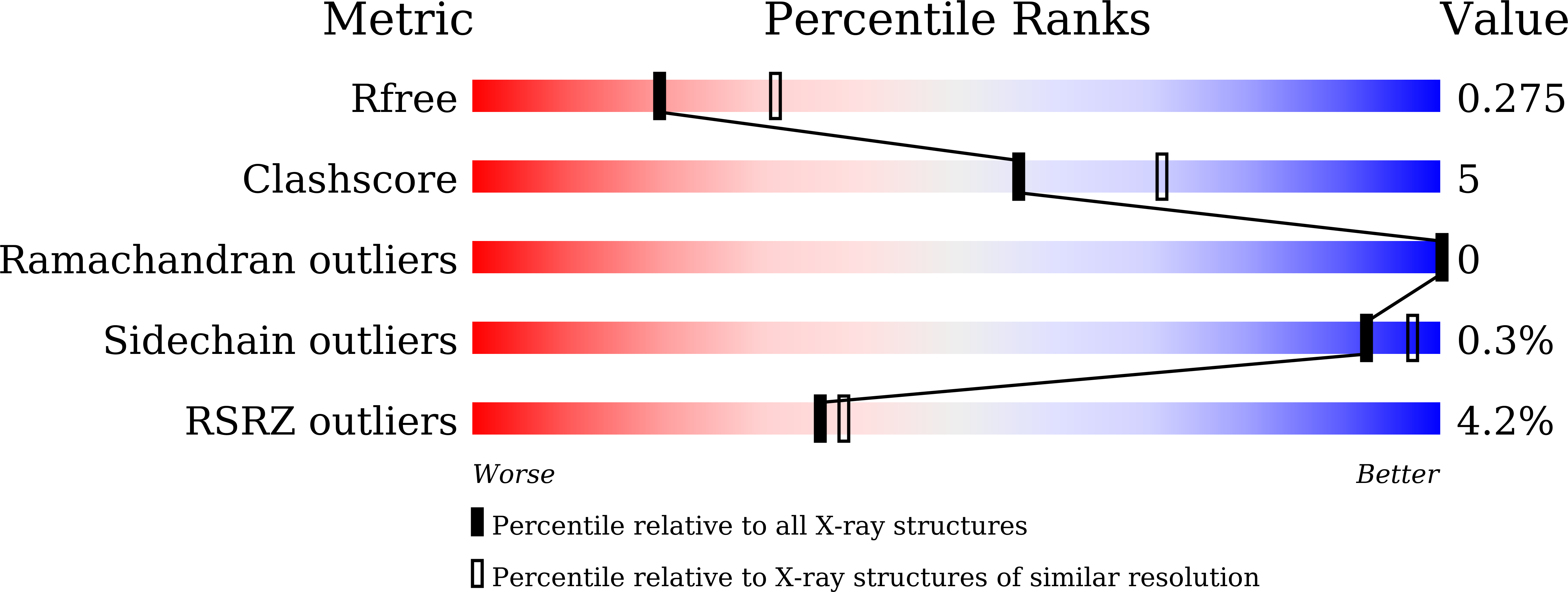
wwPDB Validation 3D Report Full Report
Entity ID: 1 | |||||
|---|---|---|---|---|---|
| Molecule | Chains | Sequence Length | Organism | Details | Image |
| Serine protease NS3 | 445 | Langat virus (strain TP21) | Mutation(s): 0 EC: 3.4.21.91 (PDB Primary Data), 3.6.1.15 (PDB Primary Data), 3.6.4.13 (PDB Primary Data) |  | |
UniProt | |||||
Find proteins for P29837 (Langat virus (strain TP21)) Explore P29837 Go to UniProtKB: P29837 | |||||
Entity Groups | |||||
| Sequence Clusters | 30% Identity50% Identity70% Identity90% Identity95% Identity100% Identity | ||||
| UniProt Group | P29837 | ||||
Sequence AnnotationsExpand | |||||
| |||||
| Length ( Å ) | Angle ( ˚ ) |
|---|---|
| a = 68.018 | α = 90 |
| b = 68.018 | β = 90 |
| c = 213.125 | γ = 90 |
| Software Name | Purpose |
|---|---|
| PHENIX | refinement |
| PHENIX | refinement |
| XDS | data reduction |
| XDS | data scaling |
| PHENIX | phasing |
| Funding Organization | Location | Grant Number |
|---|---|---|
| Other government | China | 21JCQNJC01890 |
RCSB PDB (citation) is hosted by
RCSB PDB is a member of the

