


The Crystal Structure of TTBK1 from Biortus.
Wang, F., Cheng, W., Yuan, Z., Lin, D., Ni, C.To be published.
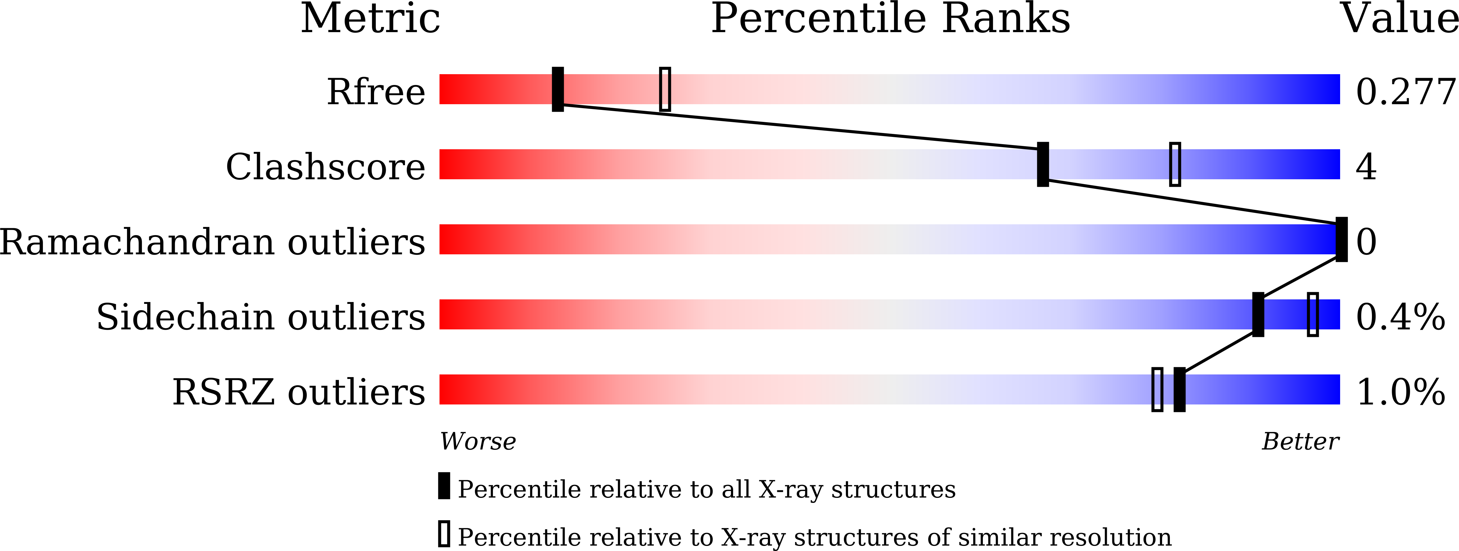
Experimental Data Snapshot
Entity ID: 1 | |||||
|---|---|---|---|---|---|
| Molecule | Chains | Sequence Length | Organism | Details | Image |
| Tau-tubulin kinase 1 | 332 | Homo sapiens | Mutation(s): 0 Gene Names: TTBK1, BDTK, KIAA1855 EC: 2.7.11.1 | 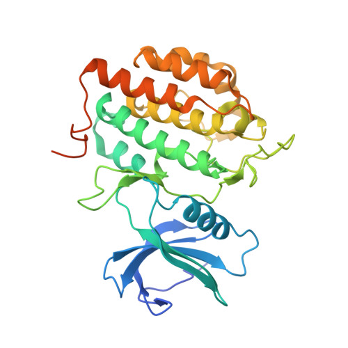 | |
UniProt & NIH Common Fund Data Resources | |||||
Find proteins for Q5TCY1 (Homo sapiens) Explore Q5TCY1 Go to UniProtKB: Q5TCY1 | |||||
PHAROS: Q5TCY1 GTEx: ENSG00000146216 | |||||
Entity Groups | |||||
| Sequence Clusters | 30% Identity50% Identity70% Identity90% Identity95% Identity100% Identity | ||||
| UniProt Group | Q5TCY1 | ||||
Sequence AnnotationsExpand | |||||
| |||||
| Ligands 1 Unique | |||||
|---|---|---|---|---|---|
| ID | Chains | Name / Formula / InChI Key | 2D Diagram | 3D Interactions | |
| EDO (Subject of Investigation/LOI) Query on EDO | B [auth A], C [auth A] | 1,2-ETHANEDIOL C2 H6 O2 LYCAIKOWRPUZTN-UHFFFAOYSA-N |  | ||
| Length ( Å ) | Angle ( ˚ ) |
|---|---|
| a = 170.905 | α = 90 |
| b = 40.204 | β = 103.725 |
| c = 49.533 | γ = 90 |
| Software Name | Purpose |
|---|---|
| REFMAC | refinement |
| XDS | data reduction |
| Aimless | data scaling |
| PHASER | phasing |
RCSB PDB (citation) is hosted by
RCSB PDB is a member of the

