


The Crystal Structure of HspBP1 from Biortus.
Wang, F., Cheng, W., Lv, Z., Meng, Q., Xu, Y.To be published.
Experimental Data Snapshot
wwPDB Validation 3D Report Full Report

Entity ID: 1 | |||||
|---|---|---|---|---|---|
| Molecule | Chains | Sequence Length | Organism | Details | Image |
| Hsp70-binding protein 1 | 276 | Homo sapiens | Mutation(s): 1 Gene Names: HSPBP1 | 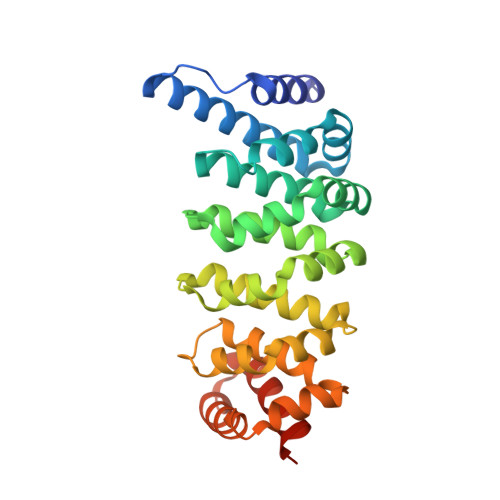 | |
UniProt & NIH Common Fund Data Resources | |||||
Find proteins for Q9NZL4 (Homo sapiens) Explore Q9NZL4 Go to UniProtKB: Q9NZL4 | |||||
PHAROS: Q9NZL4 GTEx: ENSG00000133265 | |||||
Entity Groups | |||||
| Sequence Clusters | 30% Identity50% Identity70% Identity90% Identity95% Identity100% Identity | ||||
| UniProt Group | Q9NZL4 | ||||
Sequence AnnotationsExpand | |||||
| |||||
| Length ( Å ) | Angle ( ˚ ) |
|---|---|
| a = 76.931 | α = 90 |
| b = 89.411 | β = 90 |
| c = 84.68 | γ = 90 |
| Software Name | Purpose |
|---|---|
| REFMAC | refinement |
| XDS | data reduction |
| Aimless | data scaling |
| PHASER | phasing |
RCSB PDB (citation) is hosted by
RCSB PDB is a member of the

