8WYE

Cryo-EM structure of DSR2-DSAD1 (partial) complex


Experimental Data Snapshot

  • Method: ELECTRON MICROSCOPY
  • Resolution: 2.49 Å
  • Aggregation State: PARTICLE 
  • Reconstruction Method: SINGLE PARTICLE 

wwPDB Validation   3D Report Full Report


This is version 1.1 of the entry. See complete history


Literature

Structural basis for phage-mediated activation and repression of bacterial DSR2 anti-phage defense system.

Zhang, J.T.Liu, X.Y.Li, Z.Wei, X.Y.Song, X.Y.Cui, N.Zhong, J.Li, H.Jia, N.

(2024) Nat Commun 15: 2797-2797

  • DOI: https://doi.org/10.1038/s41467-024-47177-9
  • Primary Citation of Related Structures:  
    8WY8, 8WY9, 8WYA, 8WYB, 8WYC, 8WYD, 8WYE, 8WYF

  • PubMed Abstract: 

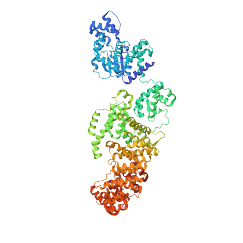
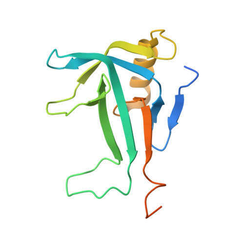
    Silent information regulator 2 (Sir2) proteins typically catalyze NAD + -dependent protein deacetylation. The recently identified bacterial Sir2 domain-containing protein, defense-associated sirtuin 2 (DSR2), recognizes the phage tail tube and depletes NAD + to abort phage propagation, which is counteracted by the phage-encoded DSR anti-defense 1 (DSAD1), but their molecular mechanisms remain unclear. Here, we determine cryo-EM structures of inactive DSR2 in its apo form, DSR2-DSAD1 and DSR2-DSAD1-NAD + , as well as active DSR2-tube and DSR2-tube-NAD + complexes. DSR2 forms a tetramer with its C-terminal sensor domains (CTDs) in two distinct conformations: CTD closed or CTD open . Monomeric, rather than oligomeric, tail tube proteins preferentially bind to CTD closed and activate Sir2 for NAD + hydrolysis. DSAD1 binding to CTD open allosterically inhibits tube binding and tube-mediated DSR2 activation. Our findings provide mechanistic insight into DSR2 assembly, tube-mediated DSR2 activation, and DSAD1-mediated inhibition and NAD + substrate catalysis in bacterial DSR2 anti-phage defense systems.


  • Organizational Affiliation

    Department of Biochemistry, School of Medicine, Southern University of Science and Technology, 518055, Shenzhen, China.


Macromolecules
Find similar proteins by:  (by identity cutoff)  |  3D Structure
Entity ID: 1
MoleculeChains Sequence LengthOrganismDetailsImage
SIR2 family protein
A, B, D, E
1,005Bacillus subtilisMutation(s): 0 
Gene Names: B4122_2870
Entity Groups  
Sequence Clusters30% Identity50% Identity70% Identity90% Identity95% Identity100% Identity
Sequence Annotations
Expand
  • Reference Sequence
Find similar proteins by:  (by identity cutoff)  |  3D Structure
Entity ID: 2
MoleculeChains Sequence LengthOrganismDetailsImage
Bacillus phage SPbeta DSAD1 protein146Bacillus phage SPBc2Mutation(s): 0 
Gene Names: yotI
UniProt
Find proteins for O64191 (Bacillus phage SPbeta)
Explore O64191 
Go to UniProtKB:  O64191
Entity Groups  
Sequence Clusters30% Identity50% Identity70% Identity90% Identity95% Identity100% Identity
UniProt GroupO64191
Sequence Annotations
Expand
  • Reference Sequence
Experimental Data & Validation

Experimental Data

  • Method: ELECTRON MICROSCOPY
  • Resolution: 2.49 Å
  • Aggregation State: PARTICLE 
  • Reconstruction Method: SINGLE PARTICLE 

Structure Validation

View Full Validation Report


Entry History & Funding Information

Deposition Data


Funding OrganizationLocationGrant Number
Not funded--

Revision History  (Full details and data files)

  • Version 1.0: 2024-04-10
    Type: Initial release
  • Version 1.1: 2024-04-17
    Changes: Data collection, Structure summary