8VJS

Cryo-EM structure of Tulane virus 9-6-17 variant capsid protein VP1 9-14-18 without DTT treatment

  • Classification: VIRUS
  • Organism(s): Tulane virus
  • Mutation(s): No 

  • Deposited: 2024-01-07 Released: 2024-02-07 
  • Deposition Author(s): Sun, C., Jiang, W.
  • Funding Organization(s): National Institutes of Health/National Institute Of Allergy and Infectious Diseases (NIH/NIAID)

Experimental Data Snapshot

  • Method: ELECTRON MICROSCOPY
  • Resolution: 2.73 Å
  • Aggregation State: PARTICLE 
  • Reconstruction Method: SINGLE PARTICLE 

wwPDB Validation   3D Report Full Report


This is version 1.0 of the entry. See complete history


Literature

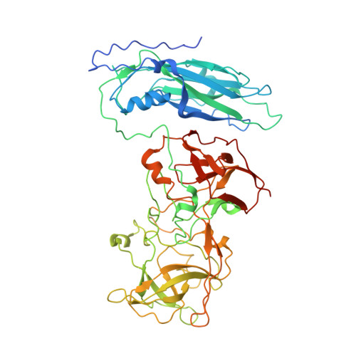
The 2.6 angstrom Structure of a Tulane Virus Variant with Minor Mutations Leading to Receptor Change.

Sun, C.Huang, P.Xu, X.Vago, F.S.Li, K.Klose, T.Jiang, X.J.Jiang, W.

(2024) Biomolecules 14

  • DOI: https://doi.org/10.3390/biom14010119
  • Primary Citation of Related Structures:  
    8VGR, 8VJR, 8VJS

  • PubMed Abstract: 

    Human noroviruses (HuNoVs) are a major cause of acute gastroenteritis, contributing significantly to annual foodborne illness cases. However, studying these viruses has been challenging due to limitations in tissue culture techniques for over four decades. Tulane virus (TV) has emerged as a crucial surrogate for HuNoVs due to its close resemblance in amino acid composition and the availability of a robust cell culture system. Initially isolated from rhesus macaques in 2008, TV represents a novel Calicivirus belonging to the Recovirus genus. Its significance lies in sharing the same host cell receptor, histo-blood group antigen (HBGA), as HuNoVs. In this study, we introduce, through cryo-electron microscopy (cryo-EM), the structure of a specific TV variant (the 9-6-17 TV) that has notably lost its ability to bind to its receptor, B-type HBGA-a finding confirmed using an enzyme-linked immunosorbent assay (ELISA). These results offer a profound insight into the genetic modifications occurring in TV that are necessary for adaptation to cell culture environments. This research significantly contributes to advancing our understanding of the genetic changes that are pivotal to successful adaptation, shedding light on fundamental aspects of Calicivirus evolution.


  • Organizational Affiliation

    Department of Biological Sciences, Purdue University, West Lafayette, IN 47907, USA.


Macromolecules
Find similar proteins by:  (by identity cutoff)  |  3D Structure
Entity ID: 1
MoleculeChains Sequence LengthOrganismDetailsImage
Capsid protein
A, B, C
534Tulane virusMutation(s): 0 
UniProt
Find proteins for B2Y6D0 (Tulane virus)
Explore B2Y6D0 
Go to UniProtKB:  B2Y6D0
Entity Groups  
Sequence Clusters30% Identity50% Identity70% Identity90% Identity95% Identity100% Identity
UniProt GroupB2Y6D0
Sequence Annotations
Expand
  • Reference Sequence
Experimental Data & Validation

Experimental Data

  • Method: ELECTRON MICROSCOPY
  • Resolution: 2.73 Å
  • Aggregation State: PARTICLE 
  • Reconstruction Method: SINGLE PARTICLE 
EM Software:
TaskSoftware PackageVersion
RECONSTRUCTIONjspr

Structure Validation

View Full Validation Report


Entry History & Funding Information

Deposition Data


Funding OrganizationLocationGrant Number
National Institutes of Health/National Institute Of Allergy and Infectious Diseases (NIH/NIAID)United StatesAI111095

Revision History  (Full details and data files)

  • Version 1.0: 2024-02-07
    Type: Initial release