8V8H

PI3Ka H1047R co-crystal structure with inhibitor in cryptic pocket near H1047R (compound 4).


Experimental Data Snapshot

  • Method: X-RAY DIFFRACTION
  • Resolution: 3.58 Å
  • R-Value Free: 0.272 
  • R-Value Work: 0.222 
  • R-Value Observed: 0.224 

wwPDB Validation   3D Report Full Report


Ligand Structure Quality Assessment 


This is version 1.2 of the entry. See complete history


Literature

Discovery of Pyridopyrimidinones that Selectively Inhibit the H1047R PI3K alpha Mutant Protein.

Ketcham, J.M.Harwood, S.J.Aranda, R.Aloiau, A.N.Bobek, B.M.Briere, D.M.Burns, A.C.Caddell Haatveit, K.Calinisan, A.Clarine, J.Elliott, A.Engstrom, L.D.Gunn, R.J.Ivetac, A.Jones, B.Kuehler, J.Lawson, J.D.Nguyen, N.Parker, C.Pearson, K.E.Rahbaek, L.Saechao, B.Wang, X.Waters, A.Waters, L.Watkins, A.H.Olson, P.Smith, C.R.Christensen, J.G.Marx, M.A.

(2024) J Med Chem 67: 4936-4949

  • DOI: https://doi.org/10.1021/acs.jmedchem.4c00078
  • Primary Citation of Related Structures:  
    8V8H, 8V8I, 8V8J, 8V8U, 8V8V

  • PubMed Abstract: 

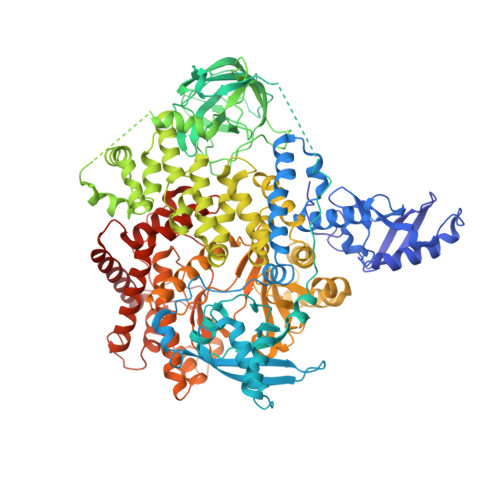
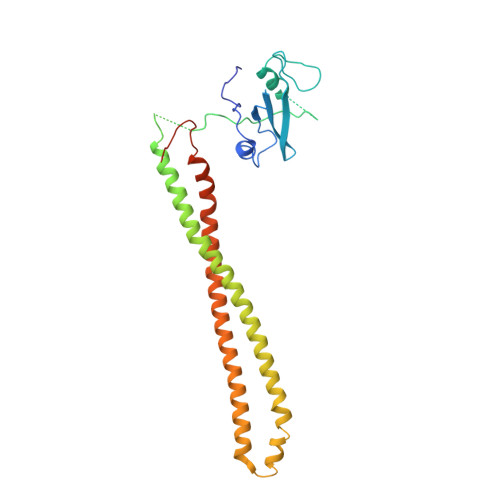
    The H1047R mutation of PIK3CA is highly prevalent in breast cancers and other solid tumors. Selectively targeting PI3Kα H1047R over PI3Kα WT is crucial due to the role that PI3Kα WT plays in normal cellular processes, including glucose homeostasis. Currently, only one PI3Kα H1047R -selective inhibitor has progressed into clinical trials, while three pan mutant (H1047R, H1047L, H1047Y, E542K, and E545K) selective PI3Kα inhibitors have also reached the clinical stage. Herein, we report the design and discovery of a series of pyridopyrimidinones that inhibit PI3Kα H1047R with high selectivity over PI3Kα WT , resulting in the discovery of compound 17 . When dosed in the HCC1954 tumor model in mice, 17 provided tumor regressions and a clear pharmacodynamic response. X-ray cocrystal structures from several PI3Kα inhibitors were obtained, revealing three distinct binding modes within PI3Kα H1047R including a previously reported cryptic pocket in the C-terminus of the kinase domain wherein we observe a ligand-induced interaction with Arg1047.


  • Organizational Affiliation

    Mirati Therapeutics, 3545 Cray Court, San Diego, California 92121, United States.


Macromolecules
Find similar proteins by:  (by identity cutoff)  |  3D Structure
Entity ID: 1
MoleculeChains Sequence LengthOrganismDetailsImage
Phosphatidylinositol 4,5-bisphosphate 3-kinase catalytic subunit alpha isoform
A, C
1,072Homo sapiensMutation(s): 1 
Gene Names: PIK3CA
UniProt & NIH Common Fund Data Resources
Find proteins for P42336 (Homo sapiens)
Explore P42336 
Go to UniProtKB:  P42336
PHAROS:  P42336
GTEx:  ENSG00000121879 
Entity Groups  
Sequence Clusters30% Identity50% Identity70% Identity90% Identity95% Identity100% Identity
UniProt GroupP42336
Sequence Annotations
Expand
  • Reference Sequence
Find similar proteins by:  (by identity cutoff)  |  3D Structure
Entity ID: 2
MoleculeChains Sequence LengthOrganismDetailsImage
Phosphatidylinositol 3-kinase regulatory subunit alpha
B, D
279Homo sapiensMutation(s): 0 
Gene Names: PIK3R1GRB1
UniProt & NIH Common Fund Data Resources
Find proteins for P27986 (Homo sapiens)
Explore P27986 
Go to UniProtKB:  P27986
PHAROS:  P27986
GTEx:  ENSG00000145675 
Entity Groups  
Sequence Clusters30% Identity50% Identity70% Identity90% Identity95% Identity100% Identity
UniProt GroupP27986
Sequence Annotations
Expand
  • Reference Sequence
Small Molecules
Ligands 2 Unique
IDChains Name / Formula / InChI Key2D Diagram3D Interactions
YO4 (Subject of Investigation/LOI)
Query on YO4

Download Ideal Coordinates CCD File 
E [auth A],
G [auth C]
2-({(1R)-1-[2-(4,4-dimethylpiperidin-1-yl)-3,6-dimethyl-4-oxo-4H-1-benzopyran-8-yl]ethyl}amino)benzoic acid
C27 H32 N2 O4
WTFFQZJMEWAKGP-GOSISDBHSA-N
YNZ (Subject of Investigation/LOI)
Query on YNZ

Download Ideal Coordinates CCD File 
F [auth A],
H [auth C]
(2S)-N~1~-[(4P)-2-tert-butyl-4'-methyl[4,5'-bi-1,3-thiazol]-2'-yl]pyrrolidine-1,2-dicarboxamide
C17 H23 N5 O2 S2
HBPXWEPKNBHKAX-NSHDSACASA-N
Experimental Data & Validation

Experimental Data

  • Method: X-RAY DIFFRACTION
  • Resolution: 3.58 Å
  • R-Value Free: 0.272 
  • R-Value Work: 0.222 
  • R-Value Observed: 0.224 
  • Space Group: C 1 2 1
Unit Cell:
Length ( Å )Angle ( ˚ )
a = 233.85α = 90
b = 157.77β = 112.66
c = 121.9γ = 90
Software Package:
Software NamePurpose
PHENIXrefinement

Structure Validation

View Full Validation Report


Ligand Structure Quality Assessment 


Entry History & Funding Information

Deposition Data


Funding OrganizationLocationGrant Number
Not fundedUnited States--

Revision History  (Full details and data files)

  • Version 1.0: 2024-03-20
    Type: Initial release
  • Version 1.1: 2024-03-27
    Changes: Database references
  • Version 1.2: 2024-04-10
    Changes: Database references