8TZO

Structure of human Wnt7a bound to WLS and CALR


Experimental Data Snapshot

  • Method: ELECTRON MICROSCOPY
  • Resolution: 3.10 Å
  • Aggregation State: PARTICLE 
  • Reconstruction Method: SINGLE PARTICLE 

wwPDB Validation   3D Report Full Report


This is version 1.1 of the entry. See complete history


Literature

Molecular basis of Wnt biogenesis, secretion, and Wnt7-specific signaling.

Qi, X.Hu, Q.Elghobashi-Meinhardt, N.Long, T.Chen, H.Li, X.

(2023) Cell 186: 5028-5040.e14

  • DOI: https://doi.org/10.1016/j.cell.2023.09.021
  • Primary Citation of Related Structures:  
    8TZO, 8TZP, 8TZS

  • PubMed Abstract: 

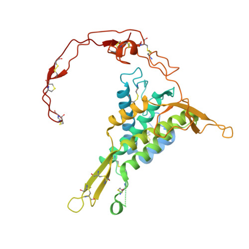
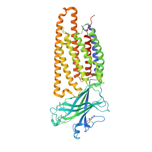
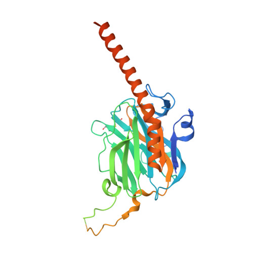
    Wnt proteins are enzymatically lipidated by Porcupine (PORCN) in the ER and bind to Wntless (WLS) for intracellular transport and secretion. Mechanisms governing the transfer of these low-solubility Wnts from the ER to the extracellular space remain unclear. Through structural and functional analyses of Wnt7a, a crucial Wnt involved in central nervous system angiogenesis and blood-brain barrier maintenance, we have elucidated the principles of Wnt biogenesis and Wnt7-specific signaling. The Wnt7a-WLS complex binds to calreticulin (CALR), revealing that CALR functions as a chaperone to facilitate Wnt transfer from PORCN to WLS during Wnt biogenesis. Our structures, functional analyses, and molecular dynamics simulations demonstrate that a phospholipid in the core of Wnt-bound WLS regulates the association and dissociation between Wnt and WLS, suggesting a lipid-mediated Wnt secretion mechanism. Finally, the structure of Wnt7a bound to RECK, a cell-surface Wnt7 co-receptor, reveals how RECK CC4 engages the N-terminal domain of Wnt7a to activate Wnt7-specific signaling.


  • Organizational Affiliation

    Department of Molecular Genetics, University of Texas Southwestern Medical Center, Dallas, TX 75390, USA. Electronic address: xiaofeng.qi@utsouthwestern.edu.


Macromolecules
Find similar proteins by:  (by identity cutoff)  |  3D Structure
Entity ID: 1
MoleculeChains Sequence LengthOrganismDetailsImage
Protein Wnt-7a349Homo sapiensMutation(s): 0 
Gene Names: WNT7A
Membrane Entity: Yes 
UniProt & NIH Common Fund Data Resources
Find proteins for O00755 (Homo sapiens)
Explore O00755 
Go to UniProtKB:  O00755
PHAROS:  O00755
GTEx:  ENSG00000154764 
Entity Groups  
Sequence Clusters30% Identity50% Identity70% Identity90% Identity95% Identity100% Identity
UniProt GroupO00755
Sequence Annotations
Expand
  • Reference Sequence
Find similar proteins by:  (by identity cutoff)  |  3D Structure
Entity ID: 2
MoleculeChains Sequence LengthOrganismDetailsImage
Protein wntless homolog541Homo sapiensMutation(s): 0 
Gene Names: WLS
Membrane Entity: Yes 
UniProt & NIH Common Fund Data Resources
Find proteins for Q5T9L3 (Homo sapiens)
Explore Q5T9L3 
Go to UniProtKB:  Q5T9L3
PHAROS:  Q5T9L3
GTEx:  ENSG00000116729 
Entity Groups  
Sequence Clusters30% Identity50% Identity70% Identity90% Identity95% Identity100% Identity
UniProt GroupQ5T9L3
Sequence Annotations
Expand
  • Reference Sequence
Find similar proteins by:  (by identity cutoff)  |  3D Structure
Entity ID: 3
MoleculeChains Sequence LengthOrganismDetailsImage
Calreticulin417Homo sapiensMutation(s): 0 
Membrane Entity: Yes 
UniProt & NIH Common Fund Data Resources
Find proteins for P27797 (Homo sapiens)
Explore P27797 
Go to UniProtKB:  P27797
PHAROS:  P27797
GTEx:  ENSG00000179218 
Entity Groups  
Sequence Clusters30% Identity50% Identity70% Identity90% Identity95% Identity100% Identity
UniProt GroupP27797
Sequence Annotations
Expand
  • Reference Sequence
Oligosaccharides

Help

Entity ID: 4
MoleculeChains Length2D Diagram Glycosylation3D Interactions
alpha-D-glucopyranose-(1-3)-alpha-D-mannopyranose-(1-2)-alpha-D-mannopyranose-(1-2)-alpha-D-mannopyranose-(1-3)-beta-D-mannopyranose-(1-4)-2-acetamido-2-deoxy-beta-D-glucopyranose-(1-4)-2-acetamido-2-deoxy-beta-D-glucopyranose
D
7N-Glycosylation
Glycosylation Resources
GlyTouCan:  G85146YR
GlyCosmos:  G85146YR
GlyGen:  G85146YR
Small Molecules
Experimental Data & Validation

Experimental Data

  • Method: ELECTRON MICROSCOPY
  • Resolution: 3.10 Å
  • Aggregation State: PARTICLE 
  • Reconstruction Method: SINGLE PARTICLE 

Structure Validation

View Full Validation Report


Entry History & Funding Information

Deposition Data


Funding OrganizationLocationGrant Number
National Institutes of Health/National Institute of General Medical Sciences (NIH/NIGMS)United StatesGM135343
National Institutes of Health/National Institute of General Medical Sciences (NIH/NIGMS)United StatesHL160487
Welch FoundationUnited StatesI-1957

Revision History  (Full details and data files)

  • Version 1.0: 2023-10-18
    Type: Initial release
  • Version 1.1: 2024-05-08
    Changes: Database references