8TQM

Cryo-EM structure of E3 ubiquitin ligase Doa10 from Saccharomyces cerevisiae


Experimental Data Snapshot

  • Method: ELECTRON MICROSCOPY
  • Resolution: 3.20 Å
  • Aggregation State: PARTICLE 
  • Reconstruction Method: SINGLE PARTICLE 

wwPDB Validation   3D Report Full Report


This is version 1.1 of the entry. See complete history


Literature

Substrate recognition mechanism of the endoplasmic reticulum-associated ubiquitin ligase Doa10.

Wu, K.Itskanov, S.Lynch, D.L.Chen, Y.Turner, A.Gumbart, J.C.Park, E.

(2024) Nat Commun 15: 2182-2182

  • DOI: https://doi.org/10.1038/s41467-024-46409-2
  • Primary Citation of Related Structures:  
    8TQM

  • PubMed Abstract: 

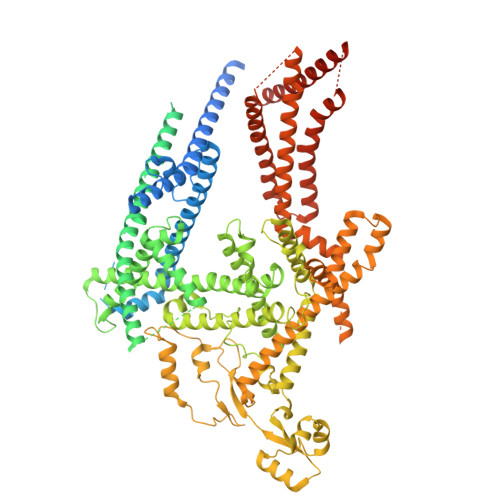
    Doa10 (MARCHF6 in metazoans) is a large polytopic membrane-embedded E3 ubiquitin ligase in the endoplasmic reticulum (ER) that plays an important role in quality control of cytosolic and ER proteins. Although Doa10 is highly conserved across eukaryotes, it is not understood how Doa10 recognizes its substrates. Here, we define the substrate recognition mechanism of Doa10 by structural and functional analyses on Saccharomyces cerevisiae Doa10 and its model substrates. Cryo-EM analysis shows that Doa10 has unusual architecture with a large lipid-filled central cavity, and its conserved middle domain forms an additional water-filled lateral tunnel open to the cytosol. Our biochemical data and molecular dynamics simulations suggest that the entrance of the substrate's degron peptide into the lateral tunnel is required for efficient polyubiquitination. The N- and C-terminal membrane domains of Doa10 seem to form fence-like features to restrict polyubiquitination to those proteins that can access the central cavity and lateral tunnel. Our study reveals how extended hydrophobic sequences at the termini of substrate proteins are recognized by Doa10 as a signal for quality control.


  • Organizational Affiliation

    Department of Molecular and Cell Biology, University of California, Berkeley, CA, 94720, USA.


Macromolecules
Find similar proteins by:  (by identity cutoff)  |  3D Structure
Entity ID: 1
MoleculeChains Sequence LengthOrganismDetailsImage
ERAD-associated E3 ubiquitin-protein ligase DOA101,319Saccharomyces cerevisiae BY4741Mutation(s): 0 
UniProt
Find proteins for P40318 (Saccharomyces cerevisiae (strain ATCC 204508 / S288c))
Explore P40318 
Go to UniProtKB:  P40318
Entity Groups  
Sequence Clusters30% Identity50% Identity70% Identity90% Identity95% Identity100% Identity
UniProt GroupP40318
Sequence Annotations
Expand
  • Reference Sequence
Small Molecules
Ligands 4 Unique
IDChains Name / Formula / InChI Key2D Diagram3D Interactions
TGL
Query on TGL

Download Ideal Coordinates CCD File 
D [auth A]TRISTEAROYLGLYCEROL
C57 H110 O6
DCXXMTOCNZCJGO-UHFFFAOYSA-N
PC1
Query on PC1

Download Ideal Coordinates CCD File 
B [auth A],
C [auth A],
E [auth A],
F [auth A]
1,2-DIACYL-SN-GLYCERO-3-PHOSPHOCHOLINE
C44 H88 N O8 P
NRJAVPSFFCBXDT-HUESYALOSA-N
Y01
Query on Y01

Download Ideal Coordinates CCD File 
G [auth A]CHOLESTEROL HEMISUCCINATE
C31 H50 O4
WLNARFZDISHUGS-MIXBDBMTSA-N
ERG
Query on ERG

Download Ideal Coordinates CCD File 
H [auth A]ERGOSTEROL
C28 H44 O
DNVPQKQSNYMLRS-APGDWVJJSA-N
Experimental Data & Validation

Experimental Data

  • Method: ELECTRON MICROSCOPY
  • Resolution: 3.20 Å
  • Aggregation State: PARTICLE 
  • Reconstruction Method: SINGLE PARTICLE 
EM Software:
TaskSoftware PackageVersion
RECONSTRUCTIONcryoSPARC3.0.0
MODEL REFINEMENTPHENIX

Structure Validation

View Full Validation Report


Entry History & Funding Information

Deposition Data


Funding OrganizationLocationGrant Number
The Vallee Foundation Inc.United States--
The Pew Charitable TrustsUnited States--

Revision History  (Full details and data files)

  • Version 1.0: 2024-03-13
    Type: Initial release
  • Version 1.1: 2024-03-27
    Changes: Database references