8T5M

SOS2 crystal structure with fragment bound (compound 14)


Experimental Data Snapshot

  • Method: X-RAY DIFFRACTION
  • Resolution: 1.79 Å
  • R-Value Free: 0.228 
  • R-Value Work: 0.190 
  • R-Value Observed: 0.192 

wwPDB Validation   3D Report Full Report


Ligand Structure Quality Assessment 


This is version 1.1 of the entry. See complete history


Literature

Discovery of Five SOS2 Fragment Hits with Binding Modes Determined by SOS2 X-Ray Cocrystallography.

Smith, C.R.Chen, D.Christensen, J.G.Coulombe, R.Fethiere, J.Gunn, R.J.Hollander, J.Jones, B.Ketcham, J.M.Khare, S.Kuehler, J.Lawson, J.D.Marx, M.A.Olson, P.Pearson, K.E.Ren, C.Tsagris, D.Ulaganathan, T.Van't Veer, I.Wang, X.Ivetac, A.

(2024) J Med Chem 67: 774-781

  • DOI: https://doi.org/10.1021/acs.jmedchem.3c02140
  • Primary Citation of Related Structures:  
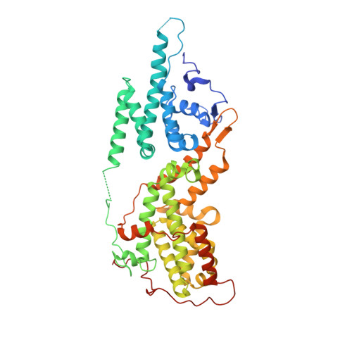
    8T5G, 8T5M, 8T5R, 8UC9, 8UF2, 8UH0

  • PubMed Abstract: 

    SOS1 and SOS2 are guanine nucleotide exchange factors that mediate RTK-stimulated RAS activation. Selective SOS1:KRAS PPI inhibitors are currently under clinical investigation, whereas there are no reports to date of SOS2:KRAS PPI inhibitors. SOS2 activity is implicated in MAPK rebound when divergent SOS1 mutant cell lines are treated with the SOS1 inhibitor BI-3406; therefore, SOS2:KRAS inhibitors are of therapeutic interest. In this report, we detail a fragment-based screening strategy to identify X-ray cocrystal structures of five diverse fragment hits bound to SOS2.


  • Organizational Affiliation

    Mirati Therapeutics, San Diego, California 92130, United States.


Macromolecules
Find similar proteins by:  (by identity cutoff)  |  3D Structure
Entity ID: 1
MoleculeChains Sequence LengthOrganismDetailsImage
Son of sevenless homolog 2490Homo sapiensMutation(s): 0 
Gene Names: SOS2
UniProt & NIH Common Fund Data Resources
Find proteins for Q07890 (Homo sapiens)
Explore Q07890 
Go to UniProtKB:  Q07890
PHAROS:  Q07890
GTEx:  ENSG00000100485 
Entity Groups  
Sequence Clusters30% Identity50% Identity70% Identity90% Identity95% Identity100% Identity
UniProt GroupQ07890
Sequence Annotations
Expand
  • Reference Sequence
Experimental Data & Validation

Experimental Data

  • Method: X-RAY DIFFRACTION
  • Resolution: 1.79 Å
  • R-Value Free: 0.228 
  • R-Value Work: 0.190 
  • R-Value Observed: 0.192 
  • Space Group: P 1
Unit Cell:
Length ( Å )Angle ( ˚ )
a = 46.471α = 85.05
b = 52.165β = 75.37
c = 60.998γ = 76.21
Software Package:
Software NamePurpose
REFMACrefinement
XDSdata reduction
XSCALEdata scaling
Cootmodel building

Structure Validation

View Full Validation Report


Ligand Structure Quality Assessment 


Entry History & Funding Information

Deposition Data


Funding OrganizationLocationGrant Number
Other privateUnited States--

Revision History  (Full details and data files)

  • Version 1.0: 2024-01-10
    Type: Initial release
  • Version 1.1: 2024-01-24
    Changes: Database references