8SYB

X-ray crystal structure of UDP-2,3-diacetamido-2,3-dideoxy-glucuronic acid-2-epimerase from Thermus thermophilus strain HB27, D98N variant in the presence of UDP-2,3-diacetamido-2,3-dideoxy-glucuronic acid and UDP-N-acetylglucosamine at pH 9


Experimental Data Snapshot

  • Method: X-RAY DIFFRACTION
  • Resolution: 2.20 Å
  • R-Value Free: 0.253 
  • R-Value Work: 0.191 
  • R-Value Observed: 0.194 

wwPDB Validation   3D Report Full Report


Ligand Structure Quality Assessment 


This is version 1.1 of the entry. See complete history


Literature

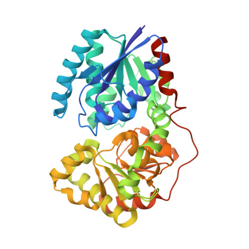
Structural analysis of a bacterial UDP-sugar 2-epimerase reveals the active site architecture before and after catalysis.

Thoden, J.B.McKnight, J.O.Kroft, C.W.Jast, J.D.T.Holden, H.M.

(2023) J Biol Chem 299: 105200-105200

  • DOI: https://doi.org/10.1016/j.jbc.2023.105200
  • Primary Citation of Related Structures:  
    8SXV, 8SXW, 8SXY, 8SY0, 8SY9, 8SYA, 8SYB, 8SYD, 8SYE, 8SYH

  • PubMed Abstract: 

    The sugar, 2,3-diacetamido-2,3-dideoxy-d-mannuronic acid, was first identified ∼40 years ago in the O-antigen of Pseudomonas aeruginosa O:3,a,d. Since then, it has been observed on the O-antigens of various pathogenic Gram-negative bacteria including Bordetella pertussis, Escherichia albertii, and Pseudomonas mediterranea. Previous studies have established that five enzymes are required for its biosynthesis beginning with uridine dinucleotide (UDP)-N-acetyl-d-glucosamine (UDP-GlcNAc). The final step in the pathway is catalyzed by a 2-epimerase, which utilizes UDP-2,3-diacetamido-2,3-dideoxy-d-glucuronic acid as its substrate. Curious as to whether this biochemical pathway is found in extreme thermophiles, we examined the published genome sequence for Thermus thermophilus HB27 and identified five ORFs that could possibly encode for the required enzymes. The focus of this investigation is on the ORF WP_011172736, which we demonstrate encodes for a 2-epimerase. For this investigation, ten high resolution X-ray crystallographic structures were determined to resolutions of 2.3 Å or higher. The models have revealed the manner in which the 2-epimerase anchors its UDP-sugar substrate as well as its UDP-sugar product into the active site. In addition, this study reveals for the first time the manner in which any sugar 2-epimerase can simultaneously bind UDP-sugars in both the active site and the allosteric binding region. We have also demonstrated that the T. thermophilus enzyme is allosterically regulated by UDP-GlcNAc. Whereas the sugar 2-epimerases that function on UDP-GlcNAc have been the focus of past biochemical and structural analyses, this is the first detailed investigation of a 2-epimerase that specifically utilizes UDP-2,3-diacetamido-2,3-dideoxy-d-glucuronic acid as its substrate.


  • Organizational Affiliation

    Department of Biochemistry, University of Wisconsin, Madison, Wisconsin, USA.


Macromolecules
Find similar proteins by:  (by identity cutoff)  |  3D Structure
Entity ID: 1
MoleculeChains Sequence LengthOrganismDetailsImage
UDP-2,3-diacetamido-2,3-dideoxy-glucuronic acid-2-epimerase
A, B
373Thermus thermophilus HB27Mutation(s): 1 
Gene Names: TT_C0285
UniProt
Find proteins for Q72KY0 (Thermus thermophilus (strain ATCC BAA-163 / DSM 7039 / HB27))
Explore Q72KY0 
Go to UniProtKB:  Q72KY0
Entity Groups  
Sequence Clusters30% Identity50% Identity70% Identity90% Identity95% Identity100% Identity
UniProt GroupQ72KY0
Sequence Annotations
Expand
  • Reference Sequence
Small Molecules
Ligands 4 Unique
IDChains Name / Formula / InChI Key2D Diagram3D Interactions
MJL (Subject of Investigation/LOI)
Query on MJL

Download Ideal Coordinates CCD File 
C [auth A],
F [auth B]
(2~{S},3~{S},4~{R},5~{R},6~{R})-4,5-diacetamido-6-[[[(2~{R},3~{S},4~{R},5~{R})-5-[2,4-bis(oxidanylidene)pyrimidin-1-yl]-3,4-bis(oxidanyl)oxolan-2-yl]methoxy-oxidanyl-phosphoryl]oxy-oxidanyl-phosphoryl]oxy-3-oxidanyl-oxane-2-carboxylic acid
C19 H28 N4 O18 P2
GZLIMKLKXDFTJR-LTMKHLKMSA-N
UD1 (Subject of Investigation/LOI)
Query on UD1

Download Ideal Coordinates CCD File 
D [auth A],
G [auth B]
URIDINE-DIPHOSPHATE-N-ACETYLGLUCOSAMINE
C17 H27 N3 O17 P2
LFTYTUAZOPRMMI-CFRASDGPSA-N
CL
Query on CL

Download Ideal Coordinates CCD File 
E [auth A],
H [auth B]
CHLORIDE ION
Cl
VEXZGXHMUGYJMC-UHFFFAOYSA-M
NA
Query on NA

Download Ideal Coordinates CCD File 
I [auth B]SODIUM ION
Na
FKNQFGJONOIPTF-UHFFFAOYSA-N
Experimental Data & Validation

Experimental Data

  • Method: X-RAY DIFFRACTION
  • Resolution: 2.20 Å
  • R-Value Free: 0.253 
  • R-Value Work: 0.191 
  • R-Value Observed: 0.194 
  • Space Group: P 1 21 1
Unit Cell:
Length ( Å )Angle ( ˚ )
a = 55.842α = 90
b = 128.195β = 113.63
c = 58.525γ = 90
Software Package:
Software NamePurpose
REFMACrefinement
SAINTdata reduction
SADABSdata scaling
PHASERphasing

Structure Validation

View Full Validation Report


Ligand Structure Quality Assessment 


Entry History & Funding Information

Deposition Data


Funding OrganizationLocationGrant Number
National Institutes of Health/National Institute of General Medical Sciences (NIH/NIGMS)United StatesR35 GM134643

Revision History  (Full details and data files)

  • Version 1.0: 2023-09-13
    Type: Initial release
  • Version 1.1: 2023-10-18
    Changes: Database references