Structural characterization of the Sel1-like repeat protein LceB from Legionella pneumophila.
Penner, T.V., Lorente Cobo, N., Patel, D.T., Patel, D.H., Savchenko, A., Brassinga, A.K.C., Prehna, G.(2024) Protein Sci 33: e4889-e4889
- PubMed: 38160319
- DOI: https://doi.org/10.1002/pro.4889
- Primary Citation of Related Structures:
8SXQ - PubMed Abstract:
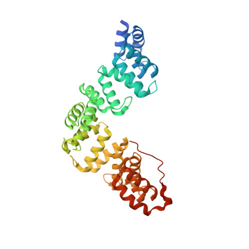
Legionella are freshwater Gram-negative bacteria that in their normal environment infect protozoa. However, this adaptation also allows Legionella to infect human alveolar macrophages and cause pneumonia. Central to Legionella pathogenesis are more than 330 secreted effectors, of which there are nine core effectors that are conserved in all pathogenic species. Despite their importance, the biochemical function of several core effectors remains unclear. To address this, we have taken a structural approach to characterize the core effector of unknown function LceB, or Lpg1356, from Legionella pneumophila. Here, we solve an X-ray crystal structure of LceB using an AlphaFold model for molecular replacement. The experimental structure shows that LceB adopts a Sel1-like repeat (SLR) fold as predicted. However, the crystal structure captured multiple conformations of LceB, all of which differed from the AlphaFold model. A comparison of the predicted model and the experimental models suggests that LceB is highly flexible in solution. Additionally, the molecular analysis of LceB using its close structural homologs reveals sequence and structural motifs of known biochemical function. Specifically, LceB harbors a repeated KAAEQG motif that both stabilizes the SLR fold and is known to participate in protein-protein interactions with eukaryotic host proteins. We also observe that LceB forms several higher-order oligomers in solution. Overall, our results have revealed that LceB has conformational flexibility, self-associates, and contains a molecular surface for binding a target host-cell protein. Additionally, our data provides structural insights into the SLR family of proteins that remain poorly studied.
Organizational Affiliation:
Department of Microbiology, University of Manitoba, Winnipeg, Manitoba, Canada.