Natural protein engineering in the Omega-loop: the role of Y221 in ceftazidime and ceftolozane resistance in Pseudomonas -derived cephalosporinase.
Mack, A.R., Kumar, V., Taracila, M.A., Mojica, M.F., O'Shea, M., Schinabeck, W., Silver, G., Hujer, A.M., Papp-Wallace, K.M., Chen, S., Haider, S., Caselli, E., Prati, F., van den Akker, F., Bonomo, R.A.(2023) Antimicrob Agents Chemother 67: e0079123-e0079123
- PubMed: 37850746
- DOI: https://doi.org/10.1128/aac.00791-23
- Primary Citation of Related Structures:
8SDL, 8SDN, 8SDR, 8SDS, 8SDT, 8SDV - PubMed Abstract:
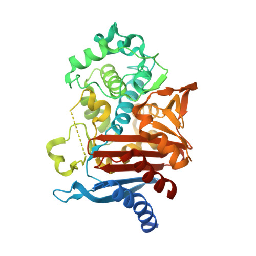
A wide variety of clinically observed single amino acid substitutions in the Ω-loop region have been associated with increased minimum inhibitory concentrations and resistance to ceftazidime (CAZ) and ceftolozane (TOL) in Pseudomonas -derived cephalosporinase and other class C β-lactamases. Herein, we demonstrate the naturally occurring tyrosine to histidine substitution of amino acid 221 (Y221H) in Pseudomonas -derived cephalosporinase (PDC) enables CAZ and TOL hydrolysis, leading to similar kinetic profiles ( k cat = 2.3 ± 0.2 µM and 2.6 ± 0.1 µM, respectively). Mass spectrometry of PDC-3 establishes the formation of stable adducts consistent with the formation of an acyl enzyme complex, while spectra of E219K (a well-characterized, CAZ- and TOL-resistant comparator) and Y221H are consistent with more rapid turnover. Thermal denaturation experiments reveal decreased stability of the variants. Importantly, PDC-3, E219K, and Y221H are all inhibited by avibactam and the boronic acid transition state inhibitors (BATSIs) LP06 and S02030 with nanomolar IC 50 values and the BATSIs stabilize all three enzymes. Crystal structures of PDC-3 and Y221H as apo enzymes and complexed with LP06 and S02030 (1.35-2.10 Å resolution) demonstrate ligand-induced conformational changes, including a significant shift in the position of the sidechain of residue 221 in Y221H (as predicted by enhanced sampling well-tempered metadynamics simulations) and extensive hydrogen bonding between the enzymes and BATSIs. The shift of residue 221 leads to the expansion of the active site pocket, and molecular docking suggests substrates orientate differently and make different intermolecular interactions in the enlarged active site compared to the wild-type enzyme.
Organizational Affiliation:
Department of Molecular Biology and Microbiology, Case Western Reserve University School of Medicine , Cleveland, Ohio, USA.