Exploring the coordination chemistry of ruthenium complexes with lysozymes: structural and in-solution studies.
Oszajca, M., Flejszar, M., Szura, A., Drozdz, P., Brindell, M., Kurpiewska, K.(2024) Front Chem 12: 1371637-1371637
- PubMed: 38638879
- DOI: https://doi.org/10.3389/fchem.2024.1371637
- Primary Citation of Related Structures:
8RNV, 8RNW, 8RNX, 8RNY - PubMed Abstract:
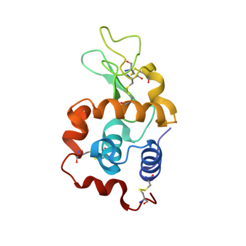
This study presents a comprehensive structural analysis of the adducts formed upon the reaction of two Ru(III) complexes [HIsq][ trans -Ru III Cl 4 (dmso)(Isq)] ( 1 ) and [H 2 Ind][ trans -Ru III Cl 4 (dmso)(HInd)] ( 2 ) (where HInd-indazole, Isq-isoquinoline, analogs of NAMI-A) and two Ru(II) complexes, cis -[RuCl 2 (dmso) 4 ] ( c ) and trans -[RuCl 2 (dmso) 4 ] ( t ), with hen-egg white lysozyme (HEWL). Additionally, the crystal structure of an adduct of human lysozyme (HL) with ruthenium complex, [H 2 Ind][ trans -RuCl 4 (dmso)(HInd)] was solved. X-ray crystallographic data analysis revealed that all studied Ru complexes, regardless of coordination surroundings and metal center charge, coordinate to the same amino acids (His15, Arg14, and Asp101) of HEWL, losing most of their original ligands. In the case of the 2 -HL adduct, two distinct metalation sites: (i) Arg107, Arg113 and (ii) Gln127, Gln129, were identified. Crystallographic data were supported by studies of the interaction of 1 and 2 with HEWL in an aqueous solution. Hydrolytic stability studies revealed that both complexes 1 and 2 liberate the N-heterocyclic ligand under crystallization-like conditions (pH 4.5) as well as under physiological pH conditions, and this process is not significantly affected by the presence of HEWL. A comparative examination of nine crystal structures of Ru complexes with lysozyme, obtained through soaking and co-crystallization experiments, together with in-solution studies of the interaction between 1 and 2 with HEWL, indicates that the hydrolytic release of the N-heterocyclic ligand is one of the critical factors in the interaction between Ru complexes and lysozyme. This understanding is crucial in shedding light on the tendency of Ru complexes to target diverse metalation sites during the formation and in the final forms of the adducts with proteins.
Organizational Affiliation:
Department of Inorganic Chemistry, Faculty of Chemistry, Jagiellonian University, Kraków, Poland.