8RIY

Human NUDT5 with ibrutinib derivative


Experimental Data Snapshot

  • Method: X-RAY DIFFRACTION
  • Resolution: 2.29 Å
  • R-Value Free: 0.295 
  • R-Value Work: 0.226 

wwPDB Validation   3D Report Full Report


Ligand Structure Quality Assessment 


This is version 1.1 of the entry. See complete history


Literature

Unexpected Noncovalent Off-Target Activity of Clinical BTK Inhibitors Leads to Discovery of a Dual NUDT5/14 Antagonist.

Balikci, E.Marques, A.M.C.Bauer, L.G.Seupel, R.Bennett, J.Raux, B.Buchan, K.Simelis, K.Singh, U.Rogers, C.Ward, J.Cheng, C.Szommer, T.Schutzenhofer, K.Elkins, J.M.Sloman, D.L.Ahel, I.Fedorov, O.Brennan, P.E.Huber, K.V.M.

(2024) J Med Chem 67: 7245-7259

  • DOI: https://doi.org/10.1021/acs.jmedchem.4c00072
  • Primary Citation of Related Structures:  
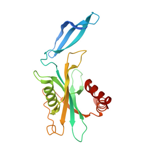
    8OTV, 8RDZ, 8RIY

  • PubMed Abstract: 

    Cofactor mimicry represents an attractive strategy for the development of enzyme inhibitors but can lead to off-target effects due to the evolutionary conservation of binding sites across the proteome. Here, we uncover the ADP-ribose (ADPr) hydrolase NUDT5 as an unexpected, noncovalent, off-target of clinical BTK inhibitors. Using a combination of biochemical, biophysical, and intact cell NanoBRET assays as well as X-ray crystallography, we confirm catalytic inhibition and cellular target engagement of NUDT5 and reveal an unusual binding mode that is independent of the reactive acrylamide warhead. Further investigation of the prototypical BTK inhibitor ibrutinib also revealed potent inhibition of the largely unstudied NUDIX hydrolase family member NUDT14. By exploring structure-activity relationships (SARs) around the core scaffold, we identify a potent, noncovalent, and cell-active dual NUDT5/14 inhibitor. Cocrystallization experiments yielded new insights into the NUDT14 hydrolase active site architecture and inhibitor binding, thus providing a basis for future chemical probe design.


  • Organizational Affiliation

    Centre for Medicines Discovery, Nuffield Department of Medicine, University of Oxford, Old Road Campus, Roosevelt Drive, Oxford OX3 7FZ, U.K.


Macromolecules
Find similar proteins by:  (by identity cutoff)  |  3D Structure
Entity ID: 1
MoleculeChains Sequence LengthOrganismDetailsImage
ADP-sugar pyrophosphataseA [auth AAA],
B [auth BBB]
209Homo sapiensMutation(s): 0 
Gene Names: NUDT5NUDIX5HSPC115
EC: 3.6.1.13 (PDB Primary Data), 3.6.1.58 (PDB Primary Data), 2.7.7.96 (PDB Primary Data)
UniProt & NIH Common Fund Data Resources
Find proteins for Q9UKK9 (Homo sapiens)
Explore Q9UKK9 
Go to UniProtKB:  Q9UKK9
PHAROS:  Q9UKK9
GTEx:  ENSG00000165609 
Entity Groups  
Sequence Clusters30% Identity50% Identity70% Identity90% Identity95% Identity100% Identity
UniProt GroupQ9UKK9
Sequence Annotations
Expand
  • Reference Sequence
Small Molecules
Ligands 1 Unique
IDChains Name / Formula / InChI Key2D Diagram3D Interactions
W0O (Subject of Investigation/LOI)
Query on W0O

Download Ideal Coordinates CCD File 
C [auth AAA],
D [auth BBB]
1-(1-methylpiperidin-4-yl)-3-(4-phenoxyphenyl)pyrazolo[3,4-d]pyrimidin-4-amine
C23 H24 N6 O
KHLKLLMMPVSSQY-UHFFFAOYSA-N
Experimental Data & Validation

Experimental Data

  • Method: X-RAY DIFFRACTION
  • Resolution: 2.29 Å
  • R-Value Free: 0.295 
  • R-Value Work: 0.226 
  • Space Group: P 61 2 2
Unit Cell:
Length ( Å )Angle ( ˚ )
a = 60.605α = 90
b = 60.605β = 90
c = 502.608γ = 120
Software Package:
Software NamePurpose
REFMACrefinement
DIALSdata reduction
Aimlessdata scaling
PHASERphasing

Structure Validation

View Full Validation Report


Ligand Structure Quality Assessment 


Entry History & Funding Information

Deposition Data


Funding OrganizationLocationGrant Number
Innovative Medicines InitiativeSwitzerland875510

Revision History  (Full details and data files)

  • Version 1.0: 2024-05-01
    Type: Initial release
  • Version 1.1: 2024-05-22
    Changes: Database references