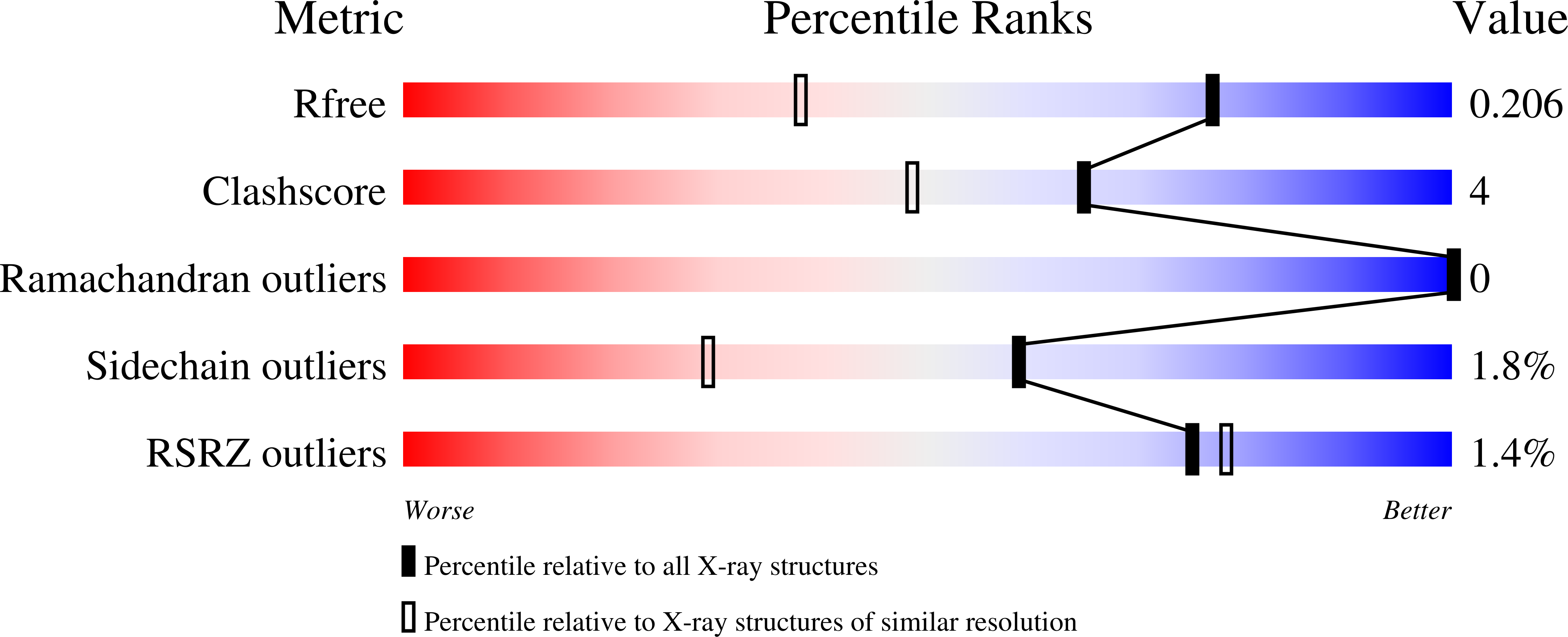
8R37

Klebsiella pneumoniae fosfomycin-resistance protein (FosAKP)


Experimental Data Snapshot

  • Method: X-RAY DIFFRACTION
  • Resolution: 1.48 Å
  • R-Value Free: 0.206 
  • R-Value Work: 0.173 
  • R-Value Observed: 0.175 

wwPDB Validation   3D Report Full Report


Ligand Structure Quality Assessment 


This is version 1.0 of the entry. See complete history


Literature

Structural Studies of Klebsiella pneumoniae Fosfomycin-Resistance Protein and Its Application for the Development of an Optical Biosensor for Fosfomycin Determination.

Varotsou, C.Ataya, F.Papageorgiou, A.C.Labrou, N.E.

(2023) Int J Mol Sci 25

  • DOI: https://doi.org/10.3390/ijms25010085
  • Primary Citation of Related Structures:  
    8R37

  • PubMed Abstract: 

    Fosfomycin-resistance proteins (FosAs) are dimeric metal-dependent glutathione transferases that conjugate the antibiotic fosfomycin (Fos) to the tripeptide glutathione (γ-Glu-Cys-Gly, GSH), rendering it inactive. In the present study, we reported a comparative analysis of the functional features of two FosAs from Pseudomonas aeruginosa (FosAPA) and Klebsiella pneumoniae (FosAKP). The coding sequences of the enzymes were cloned into a T7 expression vector, and soluble active enzymes were expressed in E. coli . FosAKP displayed higher activity and was selected for further studies. The crystal structure of the dimeric FosAKP was determined via X-ray crystallography at 1.48 Å resolution. Fos and tartrate (Tar) were found bound in the active site of the first and second molecules of the dimer, respectively. The binding of Tar to the active site caused slight rearrangements in the structure and dynamics of the enzyme, acting as a weak inhibitor of Fos binding. Differential scanning fluorimetry (DSF) was used to measure the thermal stability of FosAKP under different conditions, allowing for the selection of a suitable buffer to maximize enzyme operational stability. FosAKP displays absolute specificity towards Fos; therefore, this enzyme was exploited for the development of an enzyme-based colorimetric biosensor. FosAKP was tethered at the bottom of a plastic cuvette using glutaraldehyde chemistry to develop a simple colorimetric method for the determination of Fos in drinking water and animal plasma.


  • Organizational Affiliation

    Laboratory of Enzyme Technology, School of Applied Biology and Biotechnology, Agricultural University of Athens, 75 Iera Odos Street, GR-11855 Athens, Greece.


Macromolecules
Find similar proteins by:  (by identity cutoff)  |  3D Structure
Entity ID: 1
MoleculeChains Sequence LengthOrganismDetailsImage
FosA family fosfomycin resistance glutathione transferaseA [auth B],
B [auth A]
147Klebsiella pneumoniaeMutation(s): 0 
Gene Names: fosA
UniProt
Find proteins for A0A086IRG1 (Klebsiella pneumoniae)
Explore A0A086IRG1 
Go to UniProtKB:  A0A086IRG1
Entity Groups  
Sequence Clusters30% Identity50% Identity70% Identity90% Identity95% Identity100% Identity
UniProt GroupA0A086IRG1
Sequence Annotations
Expand
  • Reference Sequence
Small Molecules
Ligands 5 Unique
IDChains Name / Formula / InChI Key2D Diagram3D Interactions
TLA (Subject of Investigation/LOI)
Query on TLA

Download Ideal Coordinates CCD File 
M [auth A]L(+)-TARTARIC ACID
C4 H6 O6
FEWJPZIEWOKRBE-JCYAYHJZSA-N
FCN
Query on FCN

Download Ideal Coordinates CCD File 
C [auth B]FOSFOMYCIN
C3 H7 O4 P
YMDXZJFXQJVXBF-STHAYSLISA-N
MN
Query on MN

Download Ideal Coordinates CCD File 
D [auth B],
N [auth A]
MANGANESE (II) ION
Mn
WAEMQWOKJMHJLA-UHFFFAOYSA-N
K
Query on K

Download Ideal Coordinates CCD File 
L [auth B],
R [auth A]
POTASSIUM ION
K
NPYPAHLBTDXSSS-UHFFFAOYSA-N
NA
Query on NA

Download Ideal Coordinates CCD File 
E [auth B]
F [auth B]
G [auth B]
H [auth B]
I [auth B]
E [auth B],
F [auth B],
G [auth B],
H [auth B],
I [auth B],
J [auth B],
K [auth B],
O [auth A],
P [auth A],
Q [auth A]
SODIUM ION
Na
FKNQFGJONOIPTF-UHFFFAOYSA-N
Experimental Data & Validation

Experimental Data

  • Method: X-RAY DIFFRACTION
  • Resolution: 1.48 Å
  • R-Value Free: 0.206 
  • R-Value Work: 0.173 
  • R-Value Observed: 0.175 
  • Space Group: P 21 21 21
Unit Cell:
Length ( Å )Angle ( ˚ )
a = 40.62α = 90
b = 47.03β = 90
c = 149.2γ = 90
Software Package:
Software NamePurpose
PHENIXrefinement
XDSdata reduction
Aimlessdata scaling
PHASERphasing

Structure Validation

View Full Validation Report


Ligand Structure Quality Assessment 


Entry History & Funding Information

Deposition Data


Funding OrganizationLocationGrant Number
Not funded--

Revision History  (Full details and data files)

  • Version 1.0: 2024-01-24
    Type: Initial release