8R2C

Crystal structure of the Vint domain from Tetrahymena thermophila


Experimental Data Snapshot

  • Method: X-RAY DIFFRACTION
  • Resolution: 1.80 Å
  • R-Value Free: 0.226 
  • R-Value Work: 0.189 
  • R-Value Observed: 0.190 

wwPDB Validation   3D Report Full Report


This is version 1.1 of the entry. See complete history


Literature

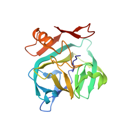
The three-dimensional structure of the Vint domain from Tetrahymena thermophila suggests a ligand-regulated cleavage mechanism by the HINT fold.

Iwai, H.Beyer, H.M.Johansson, J.E.M.Li, M.Wlodawer, A.

(2024) FEBS Lett 598: 864-874

  • DOI: https://doi.org/10.1002/1873-3468.14817
  • Primary Citation of Related Structures:  
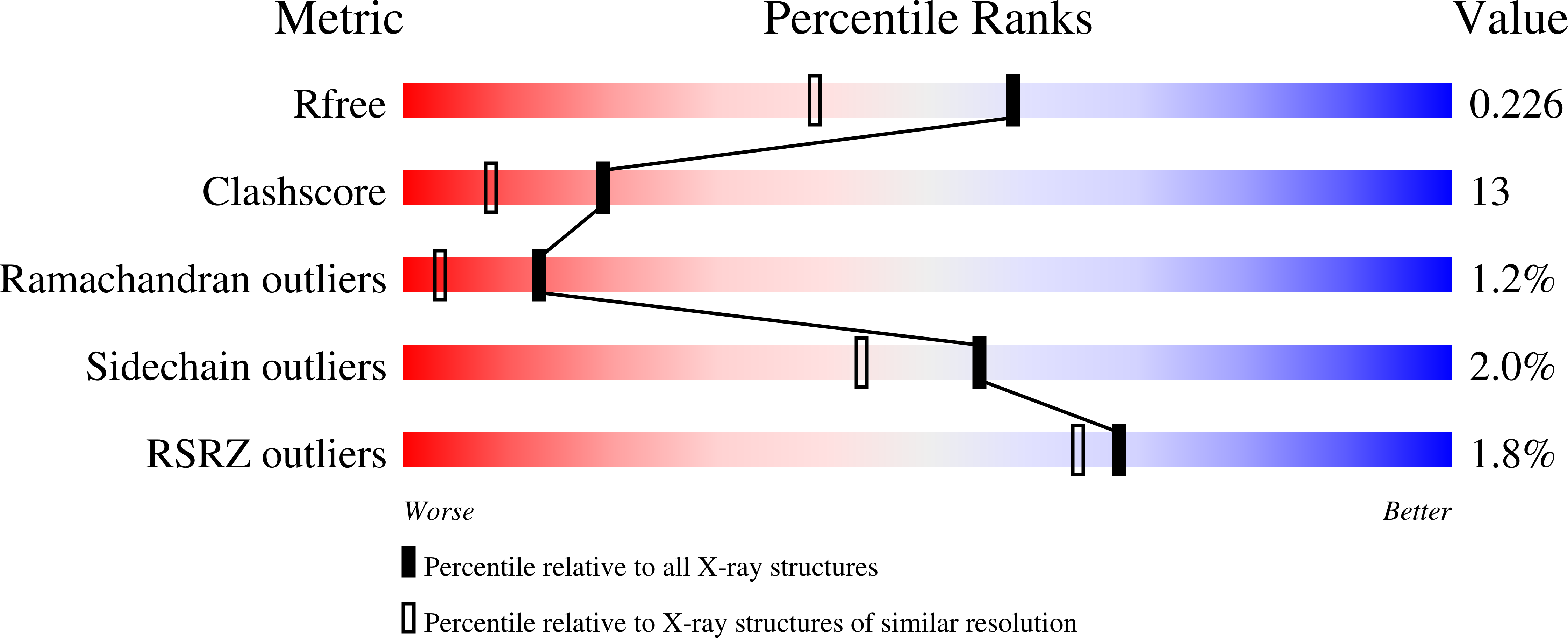
    8R2C

  • PubMed Abstract: 

    Vint proteins have been identified in unicellular metazoans as a novel hedgehog-related gene family, merging the von Willebrand factor type A domain and the Hedgehog/INTein (HINT) domains. We present the first three-dimensional structure of the Vint domain from Tetrahymena thermophila corresponding to the auto-processing domain of hedgehog proteins, shedding light on the unique features, including an adduct recognition region (ARR). Our results suggest a potential binding between the ARR and sulfated glycosaminoglycans like heparin sulfate. Moreover, we uncover a possible regulatory role of the ARR in the auto-processing by Vint domains, expanding our understanding of the HINT domain evolution and their use in biotechnological applications. Vint domains might have played a crucial role in the transition from unicellular to multicellular organisms.


  • Organizational Affiliation

    Institute of Biotechnology, University of Helsinki, Finland.


Macromolecules
Find similar proteins by:  (by identity cutoff)  |  3D Structure
Entity ID: 1
MoleculeChains Sequence LengthOrganismDetailsImage
von willebrand factor type A (VWA) domain was originally protein169Tetrahymena thermophila SB210Mutation(s): 0 
Gene Names: TTHERM_00471620
UniProt
Find proteins for I7M030 (Tetrahymena thermophila (strain SB210))
Explore I7M030 
Go to UniProtKB:  I7M030
Entity Groups  
Sequence Clusters30% Identity50% Identity70% Identity90% Identity95% Identity100% Identity
UniProt GroupI7M030
Sequence Annotations
Expand
  • Reference Sequence
Experimental Data & Validation

Experimental Data

  • Method: X-RAY DIFFRACTION
  • Resolution: 1.80 Å
  • R-Value Free: 0.226 
  • R-Value Work: 0.189 
  • R-Value Observed: 0.190 
  • Space Group: P 21 21 21
Unit Cell:
Length ( Å )Angle ( ˚ )
a = 35.89α = 90
b = 46.36β = 90
c = 115.01γ = 90
Software Package:
Software NamePurpose
PHENIXrefinement
XDSdata reduction

Structure Validation

View Full Validation Report


Entry History & Funding Information

Deposition Data


Funding OrganizationLocationGrant Number
Academy of FinlandFinland131413
Academy of FinlandFinland137995
Academy of FinlandFinland277335
Novo Nordisk FoundationDenmarkNNF17OC0025402
Novo Nordisk FoundationDenmarkNNF17OC0027550
National Institutes of Health/National Cancer Institute (NIH/NCI)United States75N91019D00024

Revision History  (Full details and data files)

  • Version 1.0: 2024-02-28
    Type: Initial release
  • Version 1.1: 2024-05-01
    Changes: Database references