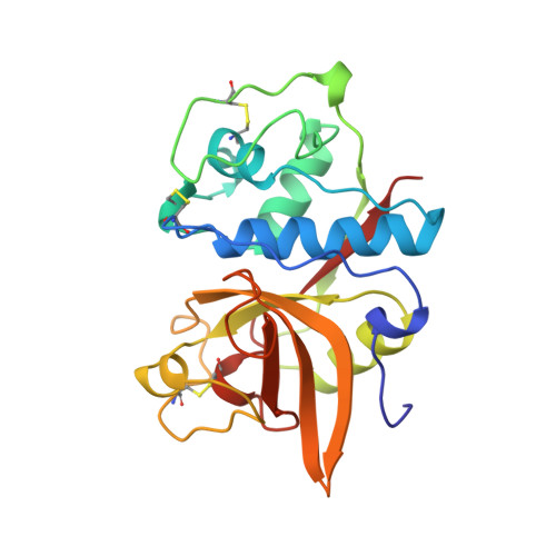
8PCH

CRYSTAL STRUCTURE OF PORCINE CATHEPSIN H DETERMINED AT 2.1 ANGSTROM RESOLUTION: LOCATION OF THE MINI-CHAIN C-TERMINAL CARBOXYL GROUP DEFINES CATHEPSIN H AMINOPEPTIDASE FUNCTION


Experimental Data Snapshot

  • Method: X-RAY DIFFRACTION
  • Resolution: 2.10 Å
  • R-Value Free: 0.245 

wwPDB Validation   3D Report Full Report


This is version 2.1 of the entry. See complete history


Literature

Crystal structure of porcine cathepsin H determined at 2.1 A resolution: location of the mini-chain C-terminal carboxyl group defines cathepsin H aminopeptidase function.

Guncar, G.Podobnik, M.Pungercar, J.Strukelj, B.Turk, V.Turk, D.

(1998) Structure 6: 51-61

  • DOI: https://doi.org/10.1016/s0969-2126(98)00007-0
  • Primary Citation of Related Structures:  
    8PCH

  • PubMed Abstract: 

    Cathepsin H is a lysosomal cysteine protease, involved in intracellular protein degradation. It is the only known mono-aminopeptidase in the papain-like family and is reported to be involved in tumor metastasis. The cathepsin H structure was determined in order to investigate the structural basis for its aminopeptidase activity and thus to provide the basis for structure-based design of synthetic inhibitors. The crystal structure of native porcine cathepsin H was determined at 2.1 A resolution. The structure has the typical papain-family fold. The so-called mini-chain, the octapeptide EPQNCSAT, is attached via a disulfide bond to the body of the enzyme and bound in a narrowed active-site cleft, in the substrate-binding direction. The mini-chain fills the region that in related enzymes comprises the non-primed substrate-binding sites from S2 backwards. The crystal structure of cathepsin H reveals that the mini-chain has a definitive role in substrate recognition and that carbohydrate residues attached to the body of the enzyme are involved in positioning the mini-chain in the active-site cleft. Modeling of a substrate into the active-site cleft suggests that the negatively charged carboxyl group of the C terminus of the mini-chain acts as an anchor for the positively charged N-terminal amino group of a substrate. The observed displacements of the residues within the active-site cleft from their equivalent positions in the papain-like endopeptidases suggest that they form the structural basis for the positioning of both the mini-chain and the substrate, resulting in exopeptidase activity.


  • Organizational Affiliation

    Department of Biochemistry and Molecular Biology, Jozef Stefan Institute, Ljubljana, Slovenia. gregor.guncar@ijs.si


Macromolecules
Find similar proteins by:  (by identity cutoff)  |  3D Structure
Entity ID: 1
MoleculeChains Sequence LengthOrganismDetailsImage
CATHEPSIN H220Sus scrofaMutation(s): 0 
EC: 3.4.22.16
UniProt
Find proteins for O46427 (Sus scrofa)
Explore O46427 
Go to UniProtKB:  O46427
Entity Groups  
Sequence Clusters30% Identity50% Identity70% Identity90% Identity95% Identity100% Identity
UniProt GroupO46427
Sequence Annotations
Expand
  • Reference Sequence

Find similar proteins by:  Sequence   |   3D Structure  

Entity ID: 2
MoleculeChains Sequence LengthOrganismDetailsImage
CATHEPSIN HB [auth P]8Sus scrofaMutation(s): 0 
EC: 3.4.22.16
UniProt
Find proteins for O46427 (Sus scrofa)
Explore O46427 
Go to UniProtKB:  O46427
Entity Groups  
UniProt GroupO46427
Sequence Annotations
Expand
  • Reference Sequence
Oligosaccharides

Help

Entity ID: 3
MoleculeChains Length2D Diagram Glycosylation3D Interactions
beta-D-mannopyranose-(1-4)-2-acetamido-2-deoxy-beta-D-glucopyranose-(1-4)-2-acetamido-2-deoxy-beta-D-glucopyranoseC [auth B]3N-Glycosylation
Glycosylation Resources
GlyTouCan:  G15407YE
GlyCosmos:  G15407YE
GlyGen:  G15407YE
Experimental Data & Validation

Experimental Data

  • Method: X-RAY DIFFRACTION
  • Resolution: 2.10 Å
  • R-Value Free: 0.245 
  • Space Group: P 21 21 21
Unit Cell:
Length ( Å )Angle ( ˚ )
a = 38.442α = 90
b = 68.73β = 90
c = 86.757γ = 90
Software Package:
Software NamePurpose
DENZOdata reduction
SCALEPACKdata scaling
MAINmodel building
MAINrefinement
MAINphasing

Structure Validation

View Full Validation Report


Entry History 

Deposition Data

Revision History  (Full details and data files)

  • Version 1.0: 1998-12-09
    Type: Initial release
  • Version 1.1: 2008-03-25
    Changes: Version format compliance
  • Version 1.2: 2011-07-13
    Changes: Non-polymer description, Version format compliance
  • Version 2.0: 2020-07-29
    Type: Remediation
    Reason: Carbohydrate remediation
    Changes: Advisory, Atomic model, Data collection, Derived calculations, Structure summary
  • Version 2.1: 2023-08-09
    Changes: Advisory, Database references, Refinement description, Structure summary