In Situ Structures of the Ultra-Long Extended and Contracted Tail of Myoviridae Phage P1.
Yang, F., Wang, L., Zhou, J., Xiao, H., Liu, H.(2023) Viruses 15
- PubMed: 37376567
- DOI: https://doi.org/10.3390/v15061267
- Primary Citation of Related Structures:
8JAJ, 8JAN - PubMed Abstract:
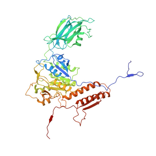
The Myoviridae phage tail is a common component of contractile injection systems (CISs), essential for exerting contractile function and facilitating membrane penetration of the inner tail tube. The near-atomic resolution structures of the Myoviridae tail have been extensively studied, but the dynamic conformational changes before and after contraction and the associated molecular mechanism are still unclear. Here, we present the extended and contracted intact tail-structures of Myoviridae phage P1 by cryo-EM. The ultra-long tail of P1, 2450 Å in length, consists of a neck, a tail terminator, 53 repeated tail sheath rings, 53 repeated tube rings, and a baseplate. The sheath of the contracted tail shrinks by approximately 55%, resulting in the separation of the inner rigid tail tube from the sheath. The extended and contracted tails were further resolved by local reconstruction at 3.3 Å and 3.9 Å resolutions, respectively, allowing us to build the atomic models of the tail terminator protein gp24, the tube protein BplB, and the sheath protein gp22 for the extended tail, and of the sheath protein gp22 for the contracted tail. Our atomic models reveal the complex interaction network in the ultra-long Myoviridae tail and the novel conformational changes of the tail sheath between extended and contracted states. Our structures provide insights into the contraction and stabilization mechanisms of the Myoviridae tail.
Organizational Affiliation:
Institute of Interdisciplinary Studies, Key Laboratory for Matter Microstructure and Function of Hunan Province, Key Laboratory of Low-Dimensional Quantum Structures and Quantum Control, Hunan Normal University, Changsha 410082, China.