


Crystal structure of a protein acetyltransferase, HP0935, acetyl-CoA bound form
Dadireddy, V., Mahanta, P., Kumar, A., Desirazu, R.N., Ramakumar, S.To be published.
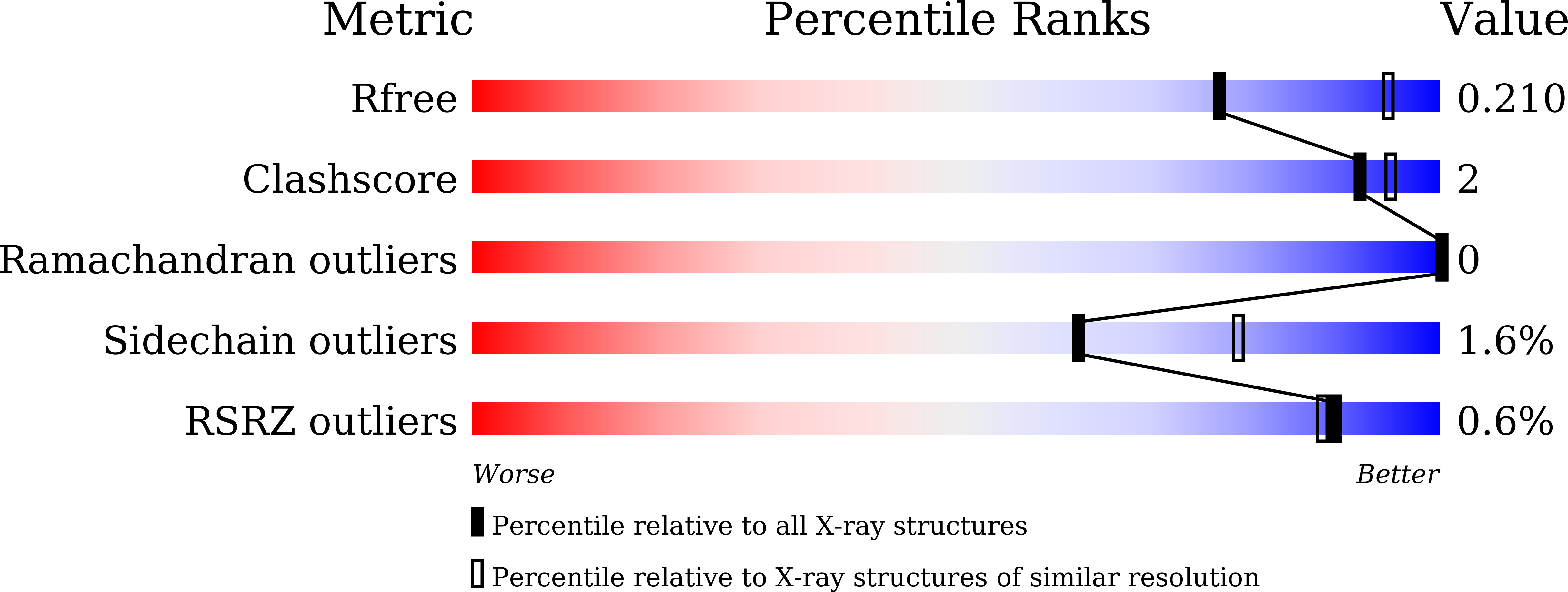
Experimental Data Snapshot
Entity ID: 1 | |||||
|---|---|---|---|---|---|
| Molecule | Chains | Sequence Length | Organism | Details | Image |
| N-acetyltransferase domain-containing protein | 163 | Helicobacter pylori 26695 | Mutation(s): 1 Gene Names: HP_0935 | 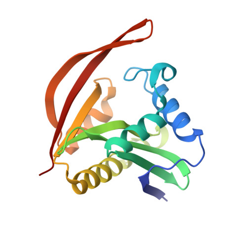 | |
UniProt | |||||
Find proteins for O25589 (Helicobacter pylori (strain ATCC 700392 / 26695)) Explore O25589 Go to UniProtKB: O25589 | |||||
Entity Groups | |||||
| Sequence Clusters | 30% Identity50% Identity70% Identity90% Identity95% Identity100% Identity | ||||
| UniProt Group | O25589 | ||||
Sequence AnnotationsExpand | |||||
| |||||
| Ligands 1 Unique | |||||
|---|---|---|---|---|---|
| ID | Chains | Name / Formula / InChI Key | 2D Diagram | 3D Interactions | |
| ACO (Subject of Investigation/LOI) Query on ACO | E [auth A], F [auth B], G [auth C], H [auth D] | ACETYL COENZYME *A C23 H38 N7 O17 P3 S ZSLZBFCDCINBPY-ZSJPKINUSA-N |  | ||
| Length ( Å ) | Angle ( ˚ ) |
|---|---|
| a = 87.797 | α = 90 |
| b = 87.797 | β = 90 |
| c = 216.36 | γ = 120 |
| Software Name | Purpose |
|---|---|
| Aimless | data scaling |
| PHASER | phasing |
| PHENIX | refinement |
| PDB_EXTRACT | data extraction |
| XDS | data reduction |
| Funding Organization | Location | Grant Number |
|---|---|---|
| Not funded | -- |
RCSB PDB (citation) is hosted by
RCSB PDB is a member of the

