


CRYSTAL STRUCTURE OF cis-N-Methylhydroxy-L-proline dehydratase in Intestinibacter bartlettii
Jiang, L., Zhang, Y.To be published.
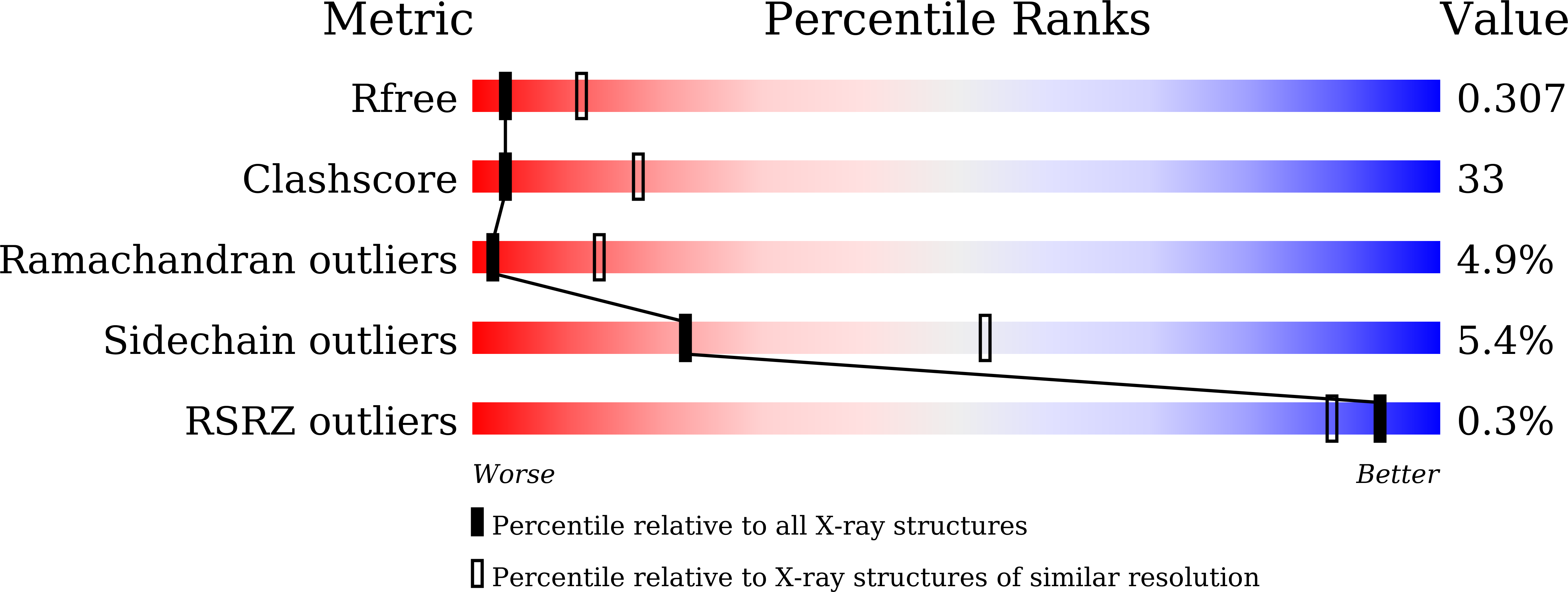
Experimental Data Snapshot
Entity ID: 1 | |||||
|---|---|---|---|---|---|
| Molecule | Chains | Sequence Length | Organism | Details | Image |
| Formate C-acetyltransferase | 792 | Intestinibacter bartlettii | Mutation(s): 0 Gene Names: C7955_105265 |  | |
Entity Groups | |||||
| Sequence Clusters | 30% Identity50% Identity70% Identity90% Identity95% Identity100% Identity | ||||
Sequence AnnotationsExpand | |||||
| |||||
| Ligands 1 Unique | |||||
|---|---|---|---|---|---|
| ID | Chains | Name / Formula / InChI Key | 2D Diagram | 3D Interactions | |
| 6IW (Subject of Investigation/LOI) Query on 6IW | B [auth A] | (2S,4S)-1-methyl-4-oxidanyl-pyrrolidine-2-carboxylic acid C6 H11 N O3 FMIPNAUMSPFTHK-WHFBIAKZSA-N |  | ||
| Length ( Å ) | Angle ( ˚ ) |
|---|---|
| a = 142.747 | α = 90 |
| b = 142.747 | β = 90 |
| c = 101.032 | γ = 90 |
| Software Name | Purpose |
|---|---|
| Aimless | data scaling |
| XDS | data reduction |
| PHENIX | refinement |
| PHENIX | phasing |
| Funding Organization | Location | Grant Number |
|---|---|---|
| National Natural Science Foundation of China (NSFC) | China | -- |
RCSB PDB (citation) is hosted by
RCSB PDB is a member of the

