8I5K

Crystal structure of chitin oligosaccharide binding protein from Vibrio cholera in complex with chitotriose.


Experimental Data Snapshot

  • Method: X-RAY DIFFRACTION
  • Resolution: 1.22 Å
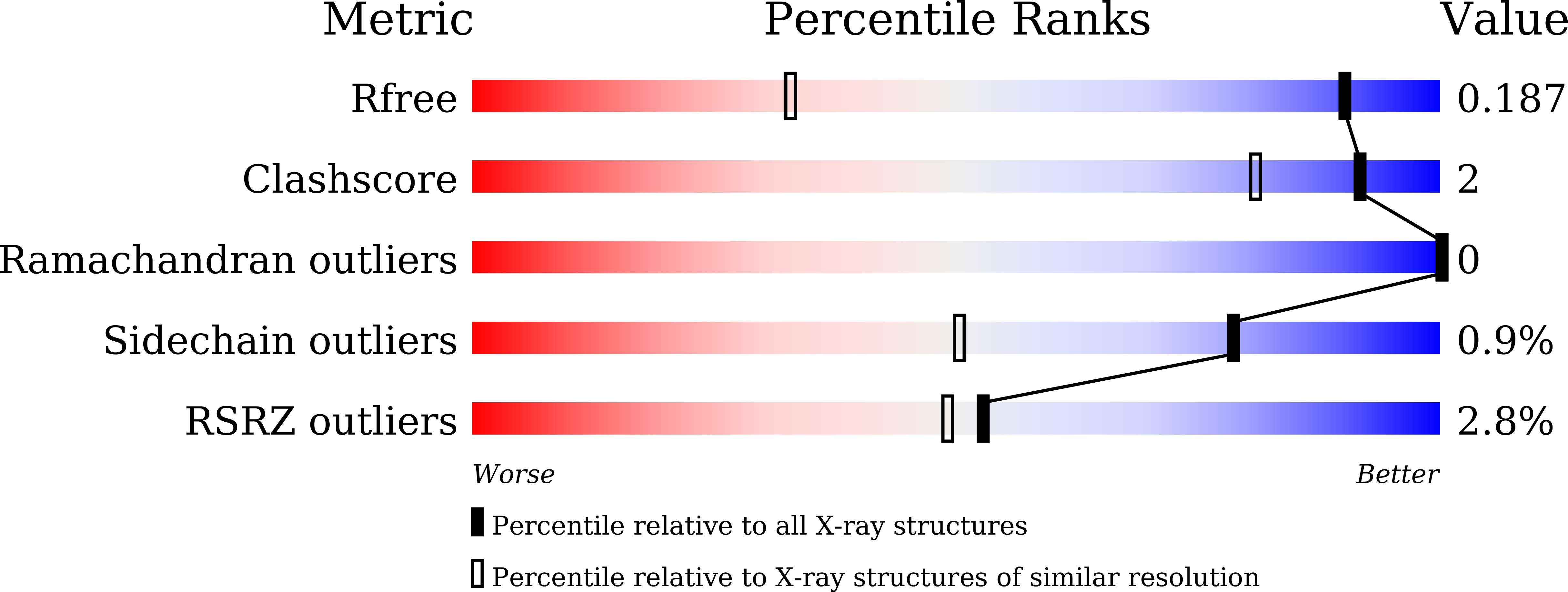
  • R-Value Free: 0.187 
  • R-Value Work: 0.169 
  • R-Value Observed: 0.170 

wwPDB Validation   3D Report Full Report


This is version 1.0 of the entry. See complete history


Literature

Periplasmic chitooligosaccharide-binding protein requires a three-domain organization for substrate translocation.

Ohnuma, T.Tsujii, J.Kataoka, C.Yoshimoto, T.Takeshita, D.Lampela, O.Juffer, A.H.Suginta, W.Fukamizo, T.

(2023) Sci Rep 13: 20558-20558

  • DOI: https://doi.org/10.1038/s41598-023-47253-y
  • Primary Citation of Related Structures:  
    8I5J, 8I5K

  • PubMed Abstract: 

    Periplasmic solute-binding proteins (SBPs) specific for chitooligosaccharides, (GlcNAc) n (n = 2, 3, 4, 5 and 6), are involved in the uptake of chitinous nutrients and the negative control of chitin signal transduction in Vibrios. Most translocation processes by SBPs across the inner membrane have been explained thus far by two-domain open/closed mechanism. Here we propose three-domain mechanism of the (GlcNAc) n translocation based on experiments using a recombinant VcCBP, SBP specific for (GlcNAc) n from Vibrio cholerae. X-ray crystal structures of unliganded or (GlcNAc) 3 -liganded VcCBP solved at 1.2-1.6 Å revealed three distinct domains, the Upper1, Upper2 and Lower domains for this protein. Molecular dynamics simulation indicated that the motions of the three domains are independent and that in the (GlcNAc) 3 -liganded state the Upper2/Lower interface fluctuated more intensively, compared to the Upper1/Lower interface. The Upper1/Lower interface bound two GlcNAc residues tightly, while the Upper2/Lower interface appeared to loosen and release the bound sugar molecule. The three-domain mechanism proposed here was fully supported by binding data obtained by thermal unfolding experiments and ITC, and may be applicable to other translocation systems involving SBPs belonging to the same cluster.


  • Organizational Affiliation

    Department of Advanced Bioscience, Kindai University, 3327-204 Nakamachi, Nara, 631-8505, Japan. ohnumat@nara.kindai.ac.jp.


Macromolecules
Find similar proteins by:  (by identity cutoff)  |  3D Structure
Entity ID: 1
MoleculeChains Sequence LengthOrganismDetailsImage
ABC transporter substrate-binding protein536Vibrio choleraeMutation(s): 0 
Gene Names: appA_1appAD6U24_16190ERS013201_01979ERS013202_02445EYB64_10090F0H40_11710F0M16_04225FLM12_17525
UniProt
Find proteins for A0A085SLP2 (Vibrio cholerae)
Explore A0A085SLP2 
Go to UniProtKB:  A0A085SLP2
Entity Groups  
Sequence Clusters30% Identity50% Identity70% Identity90% Identity95% Identity100% Identity
UniProt GroupA0A085SLP2
Sequence Annotations
Expand
  • Reference Sequence
Oligosaccharides

Help

Entity ID: 2
MoleculeChains Length2D Diagram Glycosylation3D Interactions
2-acetamido-2-deoxy-beta-D-glucopyranose-(1-4)-2-acetamido-2-deoxy-beta-D-glucopyranose-(1-4)-2-acetamido-2-deoxy-beta-D-glucopyranose
B
3N/A
Glycosylation Resources
GlyTouCan:  G47362BJ
GlyCosmos:  G47362BJ
GlyGen:  G47362BJ
Small Molecules
Ligands 1 Unique
IDChains Name / Formula / InChI Key2D Diagram3D Interactions
MG
Query on MG

Download Ideal Coordinates CCD File 
C [auth A]MAGNESIUM ION
Mg
JLVVSXFLKOJNIY-UHFFFAOYSA-N
Biologically Interesting Molecules (External Reference) 1 Unique
Experimental Data & Validation

Experimental Data

  • Method: X-RAY DIFFRACTION
  • Resolution: 1.22 Å
  • R-Value Free: 0.187 
  • R-Value Work: 0.169 
  • R-Value Observed: 0.170 
  • Space Group: C 1 2 1
Unit Cell:
Length ( Å )Angle ( ˚ )
a = 135.087α = 90
b = 51.683β = 98.83
c = 70.328γ = 90
Software Package:
Software NamePurpose
PHENIXrefinement
XDSdata reduction
XDSdata scaling
PHASERphasing

Structure Validation

View Full Validation Report


Entry History & Funding Information

Deposition Data


Funding OrganizationLocationGrant Number
Not funded--

Revision History  (Full details and data files)

  • Version 1.0: 2024-01-03
    Type: Initial release