


Crystal structure of a bright green fluorescent protein (StayGold) with single mutation (K192Y) in jellyfish Cytaeis uchidae from Biortus
Wu, J., Wang, F., Gui, W., Cheng, W., Yang, Y.To be published.
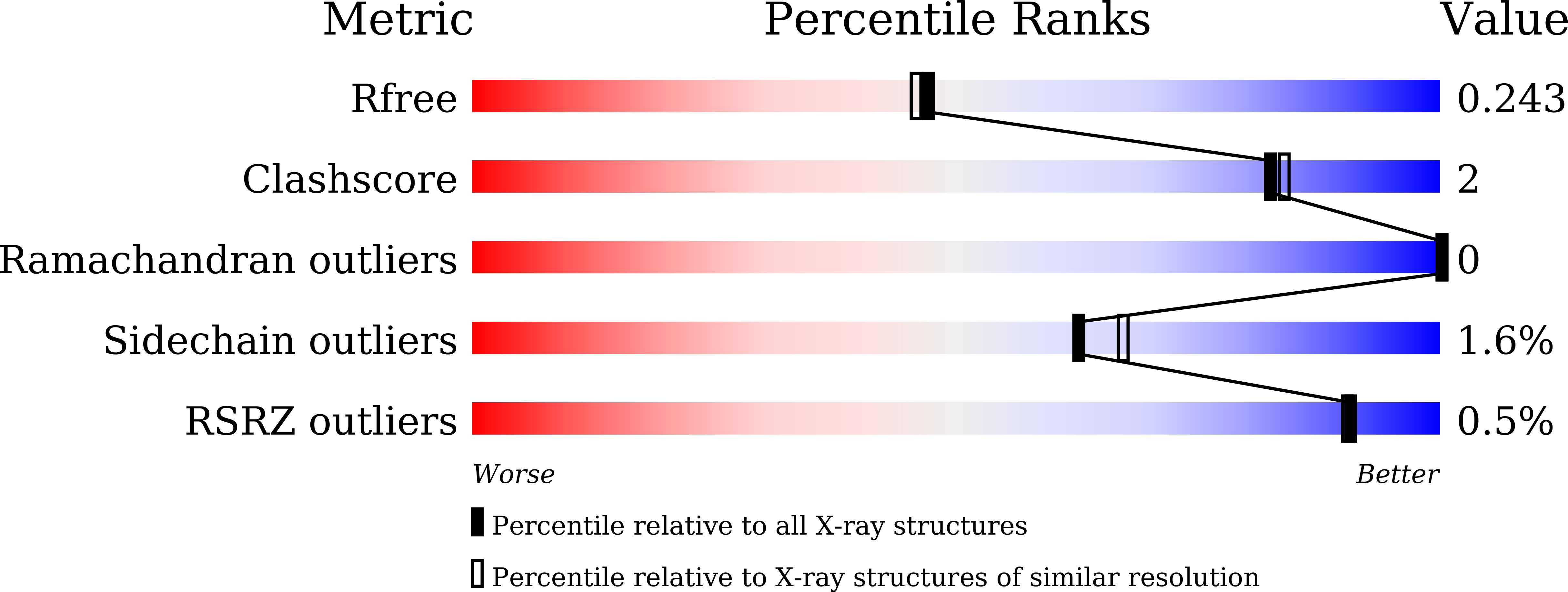
Experimental Data Snapshot
wwPDB Validation 3D Report Full Report
Entity ID: 1 | |||||
|---|---|---|---|---|---|
| Molecule | Chains | Sequence Length | Organism | Details | Image |
| staygold(K192Y) | 242 | Cytaeis uchidae | Mutation(s): 0 | 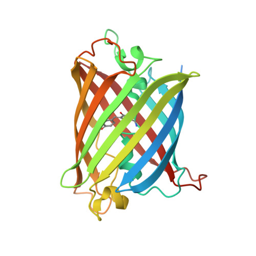 | |
UniProt | |||||
Find proteins for A0A8S0GSD4 (Cytaeis uchidae) Explore A0A8S0GSD4 Go to UniProtKB: A0A8S0GSD4 | |||||
Entity Groups | |||||
| Sequence Clusters | 30% Identity50% Identity70% Identity90% Identity95% Identity100% Identity | ||||
| UniProt Group | A0A8S0GSD4 | ||||
Sequence AnnotationsExpand | |||||
| |||||
| Ligands 1 Unique | |||||
|---|---|---|---|---|---|
| ID | Chains | Name / Formula / InChI Key | 2D Diagram | 3D Interactions | |
| EDO Query on EDO | C [auth B], D [auth B] | 1,2-ETHANEDIOL C2 H6 O2 LYCAIKOWRPUZTN-UHFFFAOYSA-N |  | ||
| Modified Residues 1 Unique | |||||
|---|---|---|---|---|---|
| ID | Chains | Type | Formula | 2D Diagram | Parent |
| CR2 Query on CR2 | A, B | L-PEPTIDE LINKING | C13 H13 N3 O4 |  | GLY, TYR, GLY |
| Length ( Å ) | Angle ( ˚ ) |
|---|---|
| a = 134.707 | α = 90 |
| b = 134.707 | β = 90 |
| c = 58.568 | γ = 120 |
| Software Name | Purpose |
|---|---|
| REFMAC | refinement |
| XDS | data reduction |
| Aimless | data scaling |
| PHASER | phasing |
RCSB PDB (citation) is hosted by
RCSB PDB is a member of the

