


Mechanism of the CBM35 domain in assisting catalysis by Ape1, a Campylobacter jejuni O-acetyl esterase
Lin, C.S., Yen, I.Y., Chan, A.C., Murphy, M.E.To be published.
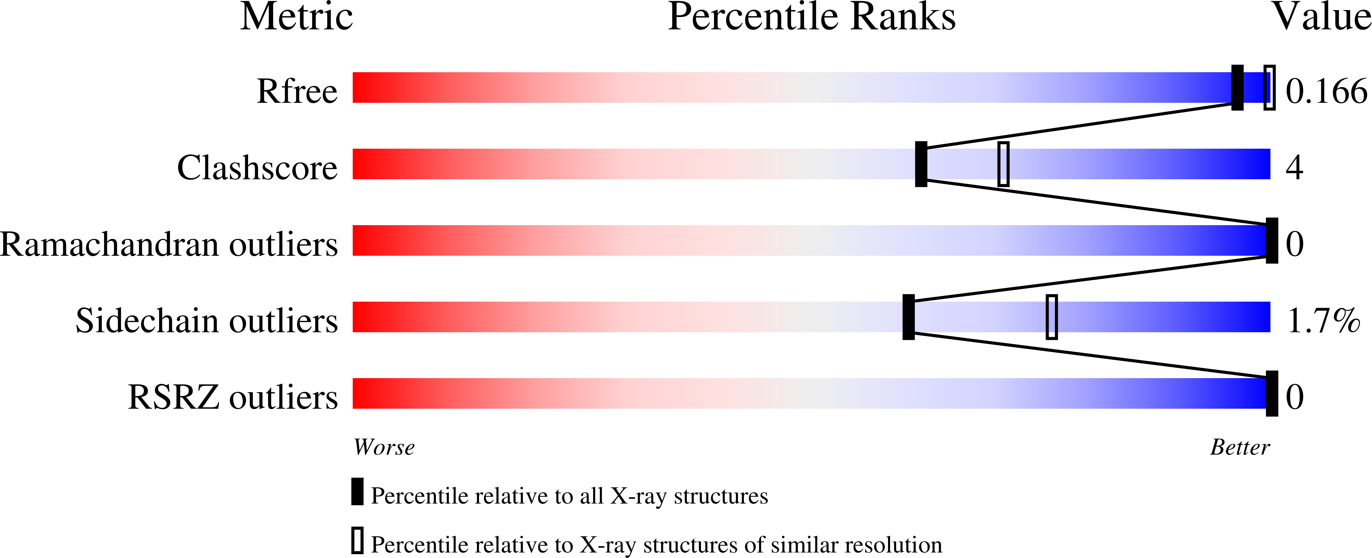
Experimental Data Snapshot
Entity ID: 1 | |||||
|---|---|---|---|---|---|
| Molecule | Chains | Sequence Length | Organism | Details | Image |
| SGNH hydrolase-type esterase domain-containing protein | 377 | Campylobacter jejuni subsp. jejuni 81-176 | Mutation(s): 0 Gene Names: CJJ81176_0638 | 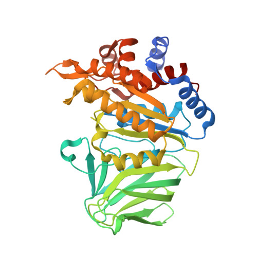 | |
UniProt | |||||
Find proteins for A0A0H3PJ52 (Campylobacter jejuni subsp. jejuni serotype O:23/36 (strain 81-176)) Explore A0A0H3PJ52 Go to UniProtKB: A0A0H3PJ52 | |||||
Entity Groups | |||||
| Sequence Clusters | 30% Identity50% Identity70% Identity90% Identity95% Identity100% Identity | ||||
| UniProt Group | A0A0H3PJ52 | ||||
Sequence AnnotationsExpand | |||||
| |||||
| Ligands 1 Unique | |||||
|---|---|---|---|---|---|
| ID | Chains | Name / Formula / InChI Key | 2D Diagram | 3D Interactions | |
| ACT (Subject of Investigation/LOI) Query on ACT | D [auth A], E [auth B], F [auth C] | ACETATE ION C2 H3 O2 QTBSBXVTEAMEQO-UHFFFAOYSA-M |  | ||
| Length ( Å ) | Angle ( ˚ ) |
|---|---|
| a = 95.311 | α = 90 |
| b = 95.311 | β = 90 |
| c = 103.034 | γ = 120 |
| Software Name | Purpose |
|---|---|
| PHENIX | refinement |
| SCALEPACK | data scaling |
| PHASER | phasing |
| PDB_EXTRACT | data extraction |
| HKL-3000 | data reduction |
| Funding Organization | Location | Grant Number |
|---|---|---|
| Canadian Institutes of Health Research (CIHR) | Canada | MOP-142176 |
RCSB PDB (citation) is hosted by
RCSB PDB is a member of the

