8FDK

Phenylhydroxylamine in Reaction with Human Hemoglobin


Experimental Data Snapshot

  • Method: X-RAY DIFFRACTION
  • Resolution: 1.89 Å
  • R-Value Free: 0.246 
  • R-Value Work: 0.208 
  • R-Value Observed: 0.210 

wwPDB Validation   3D Report Full Report


Ligand Structure Quality Assessment 


This is version 1.2 of the entry. See complete history


Literature

Crystal structural investigations of heme protein derivatives resulting from reactions of aryl- and alkylhydroxylamines with human hemoglobin.

Powell, S.M.Wang, B.Herrera, V.E.Prather, K.Y.Nguyen, N.T.Abucayon, E.G.Thomas, L.M.Safo, M.K.Richter-Addo, G.B.

(2023) J Inorg Biochem 246: 112304-112304

  • DOI: https://doi.org/10.1016/j.jinorgbio.2023.112304
  • Primary Citation of Related Structures:  
    8FDJ, 8FDK, 8FDL, 8FDM, 8FDN

  • PubMed Abstract: 

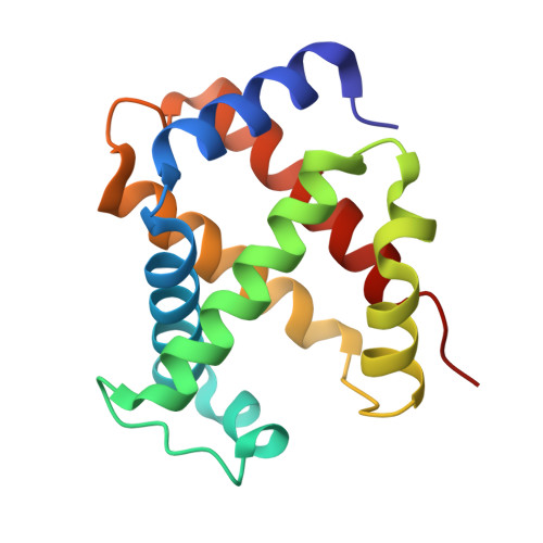
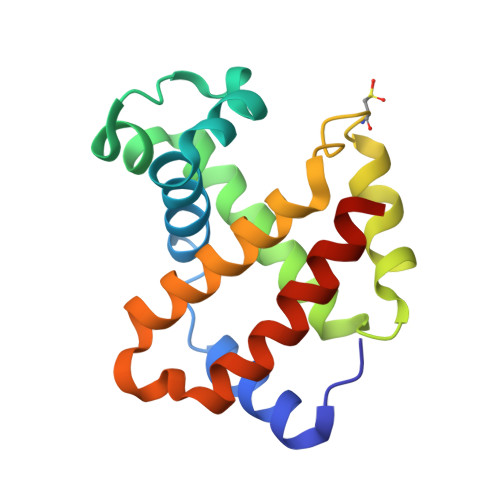
    Phenylhydroxylamine (PhNHOH) and nitrosobenzene (PhNO) interact with human tetrameric hemoglobin (Hb) to form the nitrosobenzene adduct Hb(PhNO). These interactions also frequently lead to methemoglobin formation in red blood cells. We utilize UV-vis spectroscopy and X-ray crystallography to identify the primary and secondary products that form when PhNHOH and related alkylhydroxylamines (RNHOH; R = Me, t-Bu) react with human ferric Hb. We show that with MeNHOH, the primary product is Hb[α-Fe III (H 2 O)][β-Fe II (MeNO)], in which nitrosomethane is bound to the β subunit but not the α subunit. Attempts to isolate a nitrosochloramphenicol (CAMNO) adduct resulted in our isolation of a Hb[α-Fe II ][β-Fe II -cySOx] {CAMNO} product (cySOx = oxidized cysteine) in which CAMNO was located outside of the protein in the solvent region between the β2 and α2 subunits of the same tetramer. We also observed that the βcys93 residue had been oxidized. In the case of t-BuNHOH, we demonstrate that the isolated product is the β-hemichrome Hb[α-Fe III (H 2 O)][β-Fe III (His) 2 ] {t-BuNHOH} , in which the β heme has slipped ∼4.4 Å towards the solvent exterior to accommodate the bis-His heme coordination. When PhNHOH is used, a similar β-hemichrome Hb[α-Fe III (H 2 O)][β-Fe III (His) 2 -cySOx] {PhNHOH} was obtained. Our results reveal, for the first time, the X-ray structural determination of a β-hemichrome in a human Hb derivative. Our UV-vis and X-ray crystal structural result reveal that although Hb(PhNO) and Hb(RNO) complexes may form as primary products, attempted isolation of these products by crystallization may result in the structural determination of their secondary products which may contain β-hemichromes en route to further protein degradation.


  • Organizational Affiliation

    Price Family Foundation Institute of Structural Biology, Department of Chemistry and Biochemistry, University of Oklahoma, 101 Stephenson Parkway, Norman, OK 73019, USA; Biological Sciences Division, Pacific Northwest National Laboratory, Richland, WA 99352, USA.


Macromolecules
Find similar proteins by:  (by identity cutoff)  |  3D Structure
Entity ID: 1
MoleculeChains Sequence LengthOrganismDetailsImage
Hemoglobin subunit alpha
A, C
141Homo sapiensMutation(s): 0 
UniProt & NIH Common Fund Data Resources
Find proteins for P69905 (Homo sapiens)
Explore P69905 
Go to UniProtKB:  P69905
PHAROS:  P69905
Entity Groups  
Sequence Clusters30% Identity50% Identity70% Identity90% Identity95% Identity100% Identity
UniProt GroupP69905
Sequence Annotations
Expand
  • Reference Sequence
Find similar proteins by:  (by identity cutoff)  |  3D Structure
Entity ID: 2
MoleculeChains Sequence LengthOrganismDetailsImage
Hemoglobin subunit beta
B, D
146Homo sapiensMutation(s): 0 
UniProt & NIH Common Fund Data Resources
Find proteins for P68871 (Homo sapiens)
Explore P68871 
Go to UniProtKB:  P68871
PHAROS:  P68871
GTEx:  ENSG00000244734 
Entity Groups  
Sequence Clusters30% Identity50% Identity70% Identity90% Identity95% Identity100% Identity
UniProt GroupP68871
Sequence Annotations
Expand
  • Reference Sequence
Small Molecules
Ligands 3 Unique
IDChains Name / Formula / InChI Key2D Diagram3D Interactions
HEM
Query on HEM

Download Ideal Coordinates CCD File 
E [auth A],
F [auth B],
I [auth C],
L [auth D]
PROTOPORPHYRIN IX CONTAINING FE
C34 H32 Fe N4 O4
KABFMIBPWCXCRK-RGGAHWMASA-L
NBE (Subject of Investigation/LOI)
Query on NBE

Download Ideal Coordinates CCD File 
G [auth B],
H [auth B],
J [auth C],
M [auth D],
N [auth D]
NITROSOBENZENE
C6 H5 N O
NLRKCXQQSUWLCH-UHFFFAOYSA-N
GOL
Query on GOL

Download Ideal Coordinates CCD File 
K [auth C]GLYCEROL
C3 H8 O3
PEDCQBHIVMGVHV-UHFFFAOYSA-N
Modified Residues  1 Unique
IDChains TypeFormula2D DiagramParent
CSD
Query on CSD
B, D
L-PEPTIDE LINKINGC3 H7 N O4 SCYS
Experimental Data & Validation

Experimental Data

  • Method: X-RAY DIFFRACTION
  • Resolution: 1.89 Å
  • R-Value Free: 0.246 
  • R-Value Work: 0.208 
  • R-Value Observed: 0.210 
  • Space Group: P 41
Unit Cell:
Length ( Å )Angle ( ˚ )
a = 53.263α = 90
b = 53.263β = 90
c = 194.711γ = 90
Software Package:
Software NamePurpose
PHENIXrefinement
HKL-3000data scaling
PDB_EXTRACTdata extraction
HKL-3000data collection
PHASERphasing
HKL-3000data reduction

Structure Validation

View Full Validation Report


Ligand Structure Quality Assessment 


Entry History & Funding Information

Deposition Data


Funding OrganizationLocationGrant Number
National Science Foundation (NSF, United States)United States--

Revision History  (Full details and data files)

  • Version 1.0: 2023-07-12
    Type: Initial release
  • Version 1.1: 2023-10-25
    Changes: Data collection, Refinement description
  • Version 1.2: 2024-05-01
    Changes: Database references