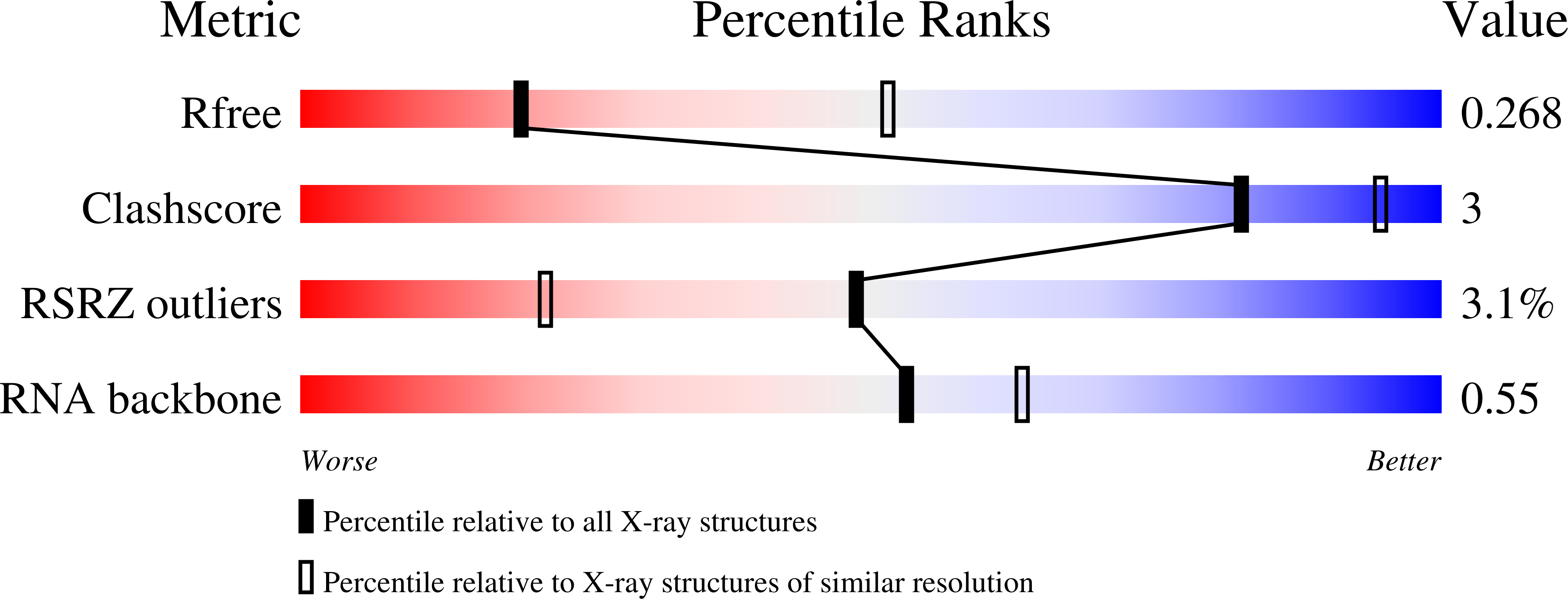
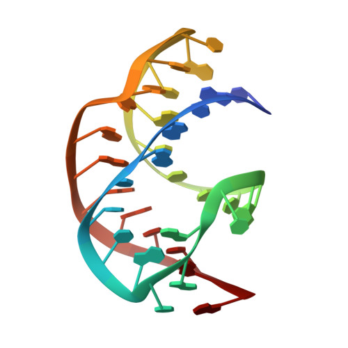
A riboswitch separated from its ribosome-binding site still regulates translation.
Schroeder, G.M., Akinyemi, O., Malik, J., Focht, C.M., Pritchett, E.M., Baker, C.D., McSally, J.P., Jenkins, J.L., Mathews, D.H., Wedekind, J.E.(2023) Nucleic Acids Res 51: 2464-2484
- PubMed: 36762498
- DOI: https://doi.org/10.1093/nar/gkad056
- Primary Citation of Related Structures:
8FB3 - PubMed Abstract:
Riboswitches regulate downstream gene expression by binding cellular metabolites. Regulation of translation initiation by riboswitches is posited to occur by metabolite-mediated sequestration of the Shine-Dalgarno sequence (SDS), causing bypass by the ribosome. Recently, we solved a co-crystal structure of a prequeuosine1-sensing riboswitch from Carnobacterium antarcticum that binds two metabolites in a single pocket. The structure revealed that the second nucleotide within the gene-regulatory SDS, G34, engages in a crystal contact, obscuring the molecular basis of gene regulation. Here, we report a co-crystal structure wherein C10 pairs with G34. However, molecular dynamics simulations reveal quick dissolution of the pair, which fails to reform. Functional and chemical probing assays inside live bacterial cells corroborate the dispensability of the C10-G34 pair in gene regulation, leading to the hypothesis that the compact pseudoknot fold is sufficient for translation attenuation. Remarkably, the C. antarcticum aptamer retained significant gene-regulatory activity when uncoupled from the SDS using unstructured spacers up to 10 nucleotides away from the riboswitch-akin to steric-blocking employed by sRNAs. Accordingly, our work reveals that the RNA fold regulates translation without SDS sequestration, expanding known riboswitch-mediated gene-regulatory mechanisms. The results infer that riboswitches exist wherein the SDS is not embedded inside a stable fold.
Organizational Affiliation:
Department of Biochemistry and Biophysics, University of Rochester School of Medicine and Dentistry, Rochester, NY 14642, USA.


