8EWY

Structure of Janus Kinase (JAK) dimer complexed with cytokine receptor intracellular domain


Experimental Data Snapshot

  • Method: ELECTRON MICROSCOPY
  • Resolution: 5.50 Å
  • Aggregation State: PARTICLE 
  • Reconstruction Method: SINGLE PARTICLE 

wwPDB Validation   3D Report Full Report


This is version 1.1 of the entry. See complete history


Literature

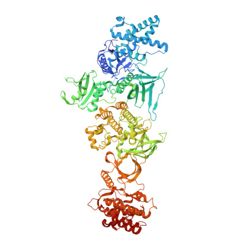
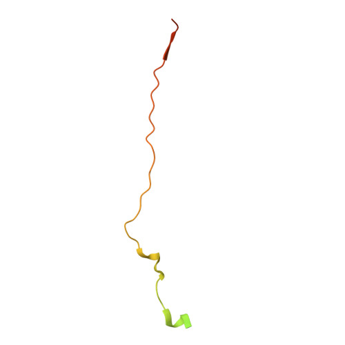
Structural basis of Janus kinase trans-activation.

Caveney, N.A.Saxton, R.A.Waghray, D.Glassman, C.R.Tsutsumi, N.Hubbard, S.R.Garcia, K.C.

(2023) Cell Rep 42: 112201-112201

  • DOI: https://doi.org/10.1016/j.celrep.2023.112201
  • Primary Citation of Related Structures:  
    8EWY

  • PubMed Abstract: 

    Janus kinases (JAKs) mediate signal transduction downstream of cytokine receptors. Cytokine-dependent dimerization is conveyed across the cell membrane to drive JAK dimerization, trans-phosphorylation, and activation. Activated JAKs in turn phosphorylate receptor intracellular domains (ICDs), resulting in the recruitment, phosphorylation, and activation of signal transducer and activator of transcription (STAT)-family transcription factors. The structural arrangement of a JAK1 dimer complex with IFNλR1 ICD was recently elucidated while bound by stabilizing nanobodies. While this revealed insights into the dimerization-dependent activation of JAKs and the role of oncogenic mutations in this process, the tyrosine kinase (TK) domains were separated by a distance not compatible with the trans-phosphorylation events between the TK domains. Here, we report the cryoelectron microscopy structure of a mouse JAK1 complex in a putative trans-activation state and expand these insights to other physiologically relevant JAK complexes, providing mechanistic insight into the crucial trans-activation step of JAK signaling and allosteric mechanisms of JAK inhibition.


  • Organizational Affiliation

    Department of Molecular and Cellular Physiology, Stanford University School of Medicine, Stanford, CA 94305, USA.


Macromolecules
Find similar proteins by:  (by identity cutoff)  |  3D Structure
Entity ID: 1
MoleculeChains Sequence LengthOrganismDetailsImage
Tyrosine-protein kinase
A, B
1,173Mus musculusMutation(s): 1 
Gene Names: Jak1
EC: 2.7.10.2
UniProt
Find proteins for P52332 (Mus musculus)
Explore P52332 
Go to UniProtKB:  P52332
Entity Groups  
Sequence Clusters30% Identity50% Identity70% Identity90% Identity95% Identity100% Identity
UniProt GroupP52332
Sequence Annotations
Expand
  • Reference Sequence
Find similar proteins by:  (by identity cutoff)  |  3D Structure
Entity ID: 2
MoleculeChains Sequence LengthOrganismDetailsImage
Interferon lambda receptor 1
C, D
85Mus musculusMutation(s): 0 
Gene Names: Ifnlr1Il28ra
UniProt & NIH Common Fund Data Resources
Find proteins for Q8CGK5 (Mus musculus)
Explore Q8CGK5 
Go to UniProtKB:  Q8CGK5
IMPC:  MGI:2429859
Entity Groups  
Sequence Clusters30% Identity50% Identity70% Identity90% Identity95% Identity100% Identity
UniProt GroupQ8CGK5
Sequence Annotations
Expand
  • Reference Sequence
Experimental Data & Validation

Experimental Data

  • Method: ELECTRON MICROSCOPY
  • Resolution: 5.50 Å
  • Aggregation State: PARTICLE 
  • Reconstruction Method: SINGLE PARTICLE 

Structure Validation

View Full Validation Report


Entry History & Funding Information

Deposition Data


Funding OrganizationLocationGrant Number
National Institutes of Health/National Institute Of Allergy and Infectious Diseases (NIH/NIAID)United StatesR37AI51321

Revision History  (Full details and data files)

  • Version 1.0: 2023-03-08
    Type: Initial release
  • Version 1.1: 2023-03-22
    Changes: Database references