Heterogeneity in M. tuberculosis beta-lactamase inhibition by Sulbactam.
Malla, T.N., Zielinski, K., Aldama, L., Bajt, S., Feliz, D., Hayes, B., Hunter, M., Kupitz, C., Lisova, S., Knoska, J., Martin-Garcia, J.M., Mariani, V., Pandey, S., Poudyal, I., Sierra, R.G., Tolstikova, A., Yefanov, O., Yoon, C.H., Ourmazd, A., Fromme, P., Schwander, P., Barty, A., Chapman, H.N., Stojkovic, E.A., Batyuk, A., Boutet, S., Phillips Jr., G.N., Pollack, L., Schmidt, M.(2023) Nat Commun 14: 5507-5507
- PubMed: 37679343
- DOI: https://doi.org/10.1038/s41467-023-41246-1
- Primary Citation of Related Structures:
8EBI, 8EBR, 8EC4, 8ECF, 8GCS, 8GCT, 8GCV, 8GCX - PubMed Abstract:
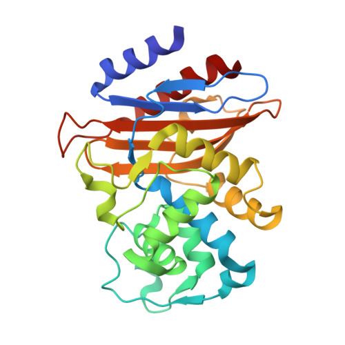
For decades, researchers have elucidated essential enzymatic functions on the atomic length scale by tracing atomic positions in real-time. Our work builds on possibilities unleashed by mix-and-inject serial crystallography (MISC) at X-ray free electron laser facilities. In this approach, enzymatic reactions are triggered by mixing substrate or ligand solutions with enzyme microcrystals. Here, we report in atomic detail (between 2.2 and 2.7 Å resolution) by room-temperature, time-resolved crystallography with millisecond time-resolution (with timepoints between 3 ms and 700 ms) how the Mycobacterium tuberculosis enzyme BlaC is inhibited by sulbactam (SUB). Our results reveal ligand binding heterogeneity, ligand gating, cooperativity, induced fit, and conformational selection all from the same set of MISC data, detailing how SUB approaches the catalytic clefts and binds to the enzyme noncovalently before reacting to a trans-enamine. This was made possible in part by the application of singular value decomposition to the MISC data using a program that remains functional even if unit cell parameters change up to 3 Å during the reaction.
Organizational Affiliation:
Physics Department, University of Wisconsin-Milwaukee, Milwaukee, WI, USA.