


Structural characterization of CMY-2 and its interactions with ampicillin and the cephalosporins - ceftiofur, DFC, DFC-dimer, DFC-cysteine, and nitrocefin
Ahmadvand, P., Call, D.R.To be published.
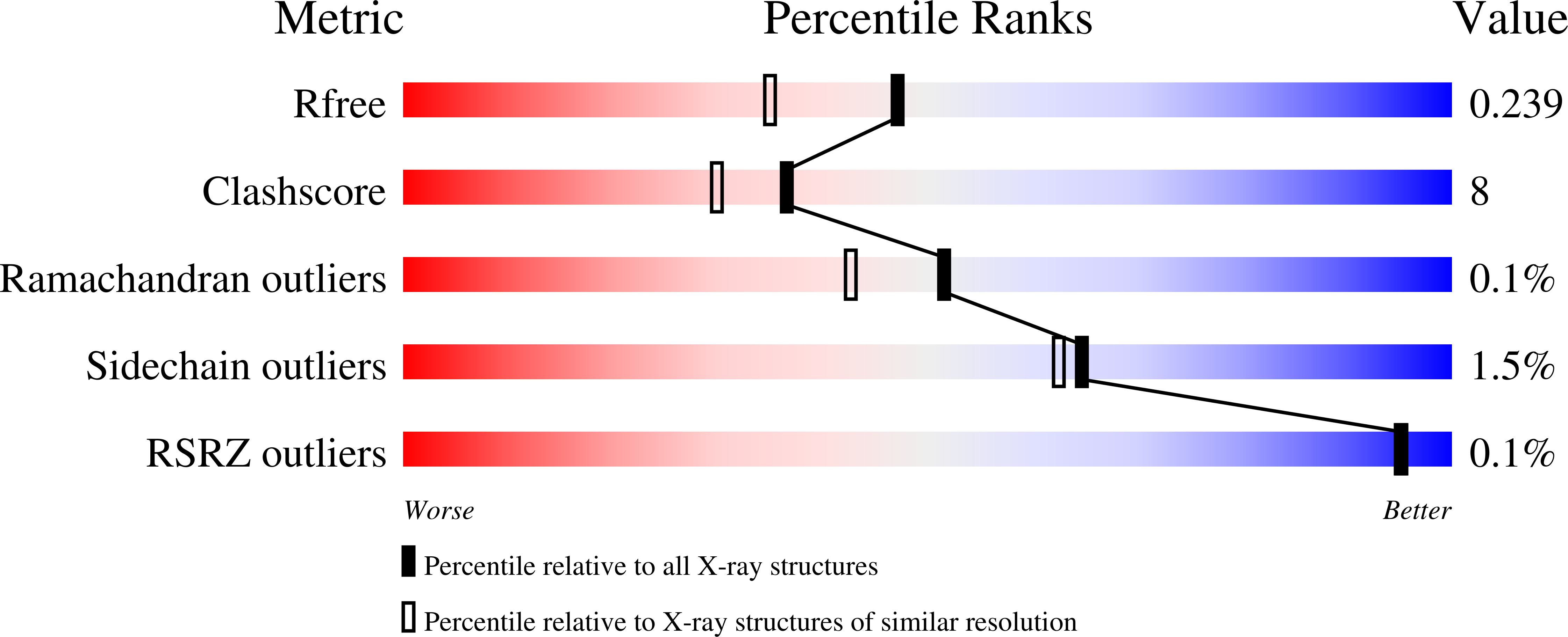
Experimental Data Snapshot
Entity ID: 1 | |||||
|---|---|---|---|---|---|
| Molecule | Chains | Sequence Length | Organism | Details | Image |
| Beta-lactamase | A [auth D], B [auth A], C [auth B], D [auth C] | 359 | Escherichia coli | Mutation(s): 0 Gene Names: blaCMY-61 EC: 3.5.2.6 | 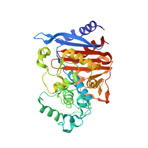 |
UniProt | |||||
Find proteins for G3F7G9 (Escherichia coli) Explore G3F7G9 Go to UniProtKB: G3F7G9 | |||||
Entity Groups | |||||
| Sequence Clusters | 30% Identity50% Identity70% Identity90% Identity95% Identity100% Identity | ||||
| UniProt Group | G3F7G9 | ||||
Sequence AnnotationsExpand | |||||
| |||||
| Ligands 1 Unique | |||||
|---|---|---|---|---|---|
| ID | Chains | Name / Formula / InChI Key | 2D Diagram | 3D Interactions | |
| SO4 (Subject of Investigation/LOI) Query on SO4 | E [auth D] F [auth D] G [auth A] H [auth A] I [auth B] | SULFATE ION O4 S QAOWNCQODCNURD-UHFFFAOYSA-L |  | ||
| Length ( Å ) | Angle ( ˚ ) |
|---|---|
| a = 55.939 | α = 81.19 |
| b = 75.374 | β = 87.84 |
| c = 97.552 | γ = 69.51 |
| Software Name | Purpose |
|---|---|
| PHENIX | refinement |
| HKL-2000 | data scaling |
| PDB_EXTRACT | data extraction |
| HKL-2000 | data reduction |
| PHENIX | phasing |
| Funding Organization | Location | Grant Number |
|---|---|---|
| National Institutes of Health/National Institute of General Medical Sciences (NIH/NIGMS) | United States | NA |
RCSB PDB (citation) is hosted by
RCSB PDB is a member of the

