8CYF

WhiB3 bound to SigmaAr4-RNAP Beta flap tip chimera and DNA


Experimental Data Snapshot

  • Method: X-RAY DIFFRACTION
  • Resolution: 2.44 Å
  • R-Value Free: 0.251 
  • R-Value Work: 0.222 
  • R-Value Observed: 0.224 

wwPDB Validation   3D Report Full Report


This is version 1.3 of the entry. See complete history


Literature

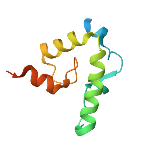
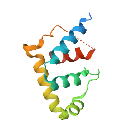
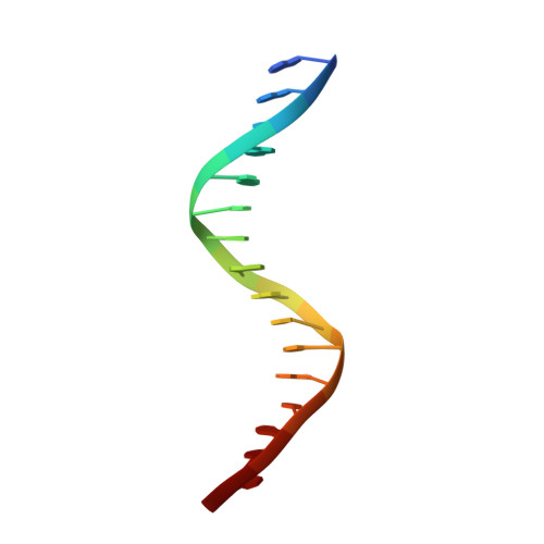
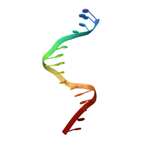
Structural basis of DNA binding by the WhiB-like transcription factor WhiB3 in Mycobacterium tuberculosis.

Wan, T.Horova, M.Khetrapal, V.Li, S.Jones, C.Schacht, A.Sun, X.Zhang, L.

(2023) J Biol Chem 299: 104777-104777

  • DOI: https://doi.org/10.1016/j.jbc.2023.104777
  • Primary Citation of Related Structures:  
    8CWR, 8CWT, 8CYF

  • PubMed Abstract: 

    Mycobacterium tuberculosis (Mtb) WhiB3 is an iron-sulfur cluster-containing transcription factor belonging to a subclass of the WhiB-Like (Wbl) family that is widely distributed in the phylum Actinobacteria. WhiB3 plays a crucial role in the survival and pathogenesis of Mtb. It binds to the conserved region 4 of the principal sigma factor (σ A 4 ) in the RNA polymerase holoenzyme to regulate gene expression like other known Wbl proteins in Mtb. However, the structural basis of how WhiB3 coordinates with σ A 4 to bind DNA and regulate transcription is unclear. Here we determined crystal structures of the WhiB3:σ A 4 complex without and with DNA at 1.5 Å and 2.45 Å, respectively, to elucidate how WhiB3 interacts with DNA to regulate gene expression. These structures reveal that the WhiB3:σ A 4 complex shares a molecular interface similar to other structurally characterized Wbl proteins and also possesses a subclass-specific Arg-rich DNA-binding motif. We demonstrate that this newly defined Arg-rich motif is required for WhiB3 binding to DNA in vitro and transcriptional regulation in Mycobacterium smegmatis. Together, our study provides empirical evidence of how WhiB3 regulates gene expression in Mtb by partnering with σ A 4 and engaging with DNA via the subclass-specific structural motif, distinct from the modes of DNA interaction by WhiB1 and WhiB7.


  • Organizational Affiliation

    Department of Biochemistry, University of Nebraska-Lincoln, Lincoln, Nebraska, USA.


Macromolecules

Find similar proteins by:  (by identity cutoff)  |  3D Structure
Entity ID: 1
MoleculeChains Sequence LengthOrganismDetailsImage
Redox- and pH-responsive transcriptional regulator WhiB3102Mycobacterium tuberculosisMutation(s): 0 
Gene Names: whiB3Rv3416
UniProt
Find proteins for P9WF41 (Mycobacterium tuberculosis (strain ATCC 25618 / H37Rv))
Explore P9WF41 
Go to UniProtKB:  P9WF41
Entity Groups  
Sequence Clusters30% Identity50% Identity70% Identity90% Identity95% Identity100% Identity
UniProt GroupP9WF41
Sequence Annotations
Expand
  • Reference Sequence
Find similar proteins by:  (by identity cutoff)  |  3D Structure
Entity ID: 2
MoleculeChains Sequence LengthOrganismDetailsImage
RNA polymerase sigma factor, DNA-directed RNA polymerase subunit beta chimera,DNA-directed RNA polymerase subunit beta112Mycobacterium tuberculosisMutation(s): 0 
Gene Names: sigAERS007670_01286ERS007681_03833ERS007688_01862
EC: 2.7.7.6
UniProt
Find proteins for P9WGY9 (Mycobacterium tuberculosis (strain ATCC 25618 / H37Rv))
Explore P9WGY9 
Go to UniProtKB:  P9WGY9
Find proteins for P9WGI1 (Mycobacterium tuberculosis (strain ATCC 25618 / H37Rv))
Explore P9WGI1 
Go to UniProtKB:  P9WGI1
Entity Groups  
Sequence Clusters30% Identity50% Identity70% Identity90% Identity95% Identity100% Identity
UniProt GroupsP9WGY9P9WGI1
Sequence Annotations
Expand
  • Reference Sequence

Find similar nucleic acids by:  Sequence   |   3D Structure  

Entity ID: 3
MoleculeChains LengthOrganismImage
DNA (5'-D(*GP*AP*AP*AP*AP*TP*CP*GP*GP*TP*TP*GP*TP*GP*GP*T)-3')16Mycobacterium tuberculosis
Sequence Annotations
Expand
  • Reference Sequence

Find similar nucleic acids by:  Sequence   |   3D Structure  

Entity ID: 4
MoleculeChains LengthOrganismImage
DNA (5'-D(*CP*AP*CP*CP*AP*CP*AP*AP*CP*CP*GP*AP*TP*TP*TP*T)-3')16Mycobacterium tuberculosis
Sequence Annotations
Expand
  • Reference Sequence
Experimental Data & Validation

Experimental Data

  • Method: X-RAY DIFFRACTION
  • Resolution: 2.44 Å
  • R-Value Free: 0.251 
  • R-Value Work: 0.222 
  • R-Value Observed: 0.224 
  • Space Group: P 21 21 21
Unit Cell:
Length ( Å )Angle ( ˚ )
a = 50.697α = 90
b = 72.934β = 90
c = 95.38γ = 90
Software Package:
Software NamePurpose
PHASERphasing
PHENIXrefinement
HKL-3000data reduction
HKL-3000data scaling
Cootmodel building

Structure Validation

View Full Validation Report


Entry History & Funding Information

Deposition Data


Funding OrganizationLocationGrant Number
National Institutes of Health/National Institute of General Medical Sciences (NIH/NIGMS)United StatesR35 GM138157-01
National Science Foundation (NSF, United States)United StatesCLP 1846908

Revision History  (Full details and data files)

  • Version 1.0: 2023-05-17
    Type: Initial release
  • Version 1.1: 2023-06-07
    Changes: Derived calculations
  • Version 1.2: 2023-06-14
    Changes: Database references
  • Version 1.3: 2023-10-25
    Changes: Data collection, Refinement description