


Membraneless channels sieve cations in ammonia-oxidizing marine archaea
von Kuegelgen, A., Cassidy, C.K., van Dorst, S., Pagani, L.L., Ford, Z., Loewe, J., Stansfeld, P.J., Bharat, T.A.M.(2024) Nature
Experimental Data Snapshot
wwPDB Validation 3D Report Full Report

(2024) Nature
Entity ID: 1 | |||||
|---|---|---|---|---|---|
| Molecule | Chains | Sequence Length | Organism | Details | Image |
| Cell surface protein | 1,734 | Nitrosopumilus maritimus SCM1 | Mutation(s): 0 | 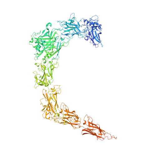 | |
UniProt | |||||
Find proteins for A9A4Y9 (Nitrosopumilus maritimus (strain SCM1)) Explore A9A4Y9 Go to UniProtKB: A9A4Y9 | |||||
Entity Groups | |||||
| Sequence Clusters | 30% Identity50% Identity70% Identity90% Identity95% Identity100% Identity | ||||
| UniProt Group | A9A4Y9 | ||||
Sequence AnnotationsExpand | |||||
| |||||
| Task | Software Package | Version |
|---|---|---|
| RECONSTRUCTION | RELION | 4.0.0 |
| MODEL REFINEMENT | PHENIX | 1.19-4092 |
| MODEL REFINEMENT | REFMAC |
| Funding Organization | Location | Grant Number |
|---|---|---|
| Wellcome Trust | United Kingdom | 202231/Z/16/Z |
| Medical Research Council (MRC, United Kingdom) | United Kingdom | MC_UP_1201/31 |
RCSB PDB (citation) is hosted by
RCSB PDB is a member of the

