8A19

Structure of a leucinostatin derivative determined by host lattice display : L1E4V1 construct


Experimental Data Snapshot

  • Method: X-RAY DIFFRACTION
  • Resolution: 2.36 Å
  • R-Value Free: 0.193 
  • R-Value Work: 0.163 
  • R-Value Observed: 0.165 

wwPDB Validation   3D Report Full Report


This is version 1.2 of the entry. See complete history


Literature

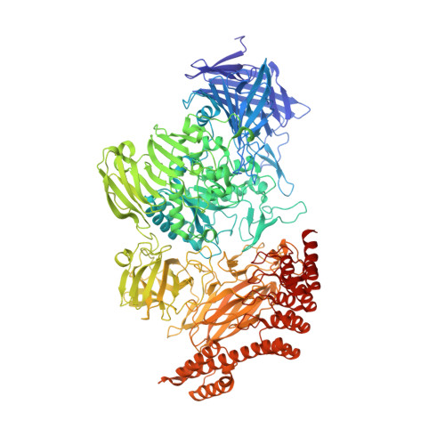
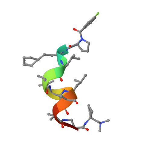
Structure of a hydrophobic leucinostatin derivative determined by host lattice display.

Kiss, C.Gall, F.M.Dreier, B.Adams, M.Riedl, R.Pluckthun, A.Mittl, P.R.E.

(2022) Acta Crystallogr D Struct Biol 78: 1439-1450

  • DOI: https://doi.org/10.1107/S2059798322010762
  • Primary Citation of Related Structures:  
    8A19, 8A1A

  • PubMed Abstract: 

    Peptides comprising many hydrophobic amino acids are almost insoluble under physiological buffer conditions, which complicates their structural analysis. To investigate the three-dimensional structure of the hydrophobic leucinostatin derivative ZHAWOC6027, the previously developed host lattice display technology was applied. Two designed ankyrin-repeat proteins (DARPins) recognizing a biotinylated ZHAWOC6027 derivative were selected from a diverse library by ribosome display under aqueous buffer conditions. ZHAWOC6027 was immobilized by means of the DARPin in the host lattice and the structure of the complex was determined by X-ray diffraction. ZHAWOC6027 adopts a distorted α-helical conformation. Comparison with the structures of related compounds that have been determined in organic solvents reveals elevated flexibility of the termini, which might be functionally important.


  • Organizational Affiliation

    Department of Biochemistry, University of Zürich, Winterthurerstrasse 190, 8057 Zürich, Switzerland.


Macromolecules
Find similar proteins by:  (by identity cutoff)  |  3D Structure
Entity ID: 1
MoleculeChains Sequence LengthOrganismDetailsImage
L1E4v11,347synthetic constructMutation(s): 0 
Entity Groups  
Sequence Clusters30% Identity50% Identity70% Identity90% Identity95% Identity100% Identity
Sequence Annotations
Expand
  • Reference Sequence

Find similar proteins by:  Sequence   |   3D Structure  

Entity ID: 2
MoleculeChains Sequence LengthOrganismDetailsImage
6-(2-methoxyethoxy)-11,15-dimethyl-8-oxa-2,11,15,19,21,23-hexazatetracyclo[15.6.1.13,7.020,24]pentacosa-1(23),3(25),4,6,17,20(24),21-heptaen-10-one11synthetic constructMutation(s): 0 
Entity Groups  
Sequence Clusters30% Identity50% Identity70% Identity90% Identity95% Identity100% Identity
Sequence Annotations
Expand
  • Reference Sequence
Small Molecules
Ligands 4 Unique
IDChains Name / Formula / InChI Key2D Diagram3D Interactions
MES
Query on MES

Download Ideal Coordinates CCD File 
K [auth A]2-(N-MORPHOLINO)-ETHANESULFONIC ACID
C6 H13 N O4 S
SXGZJKUKBWWHRA-UHFFFAOYSA-N
MPD
Query on MPD

Download Ideal Coordinates CCD File 
G [auth A],
H [auth A],
I [auth A],
J [auth A]
(4S)-2-METHYL-2,4-PENTANEDIOL
C6 H14 O2
SVTBMSDMJJWYQN-YFKPBYRVSA-N
MN
Query on MN

Download Ideal Coordinates CCD File 
C [auth A],
D [auth A],
E [auth A],
F [auth A]
MANGANESE (II) ION
Mn
WAEMQWOKJMHJLA-UHFFFAOYSA-N
CL
Query on CL

Download Ideal Coordinates CCD File 
L [auth A],
M [auth A],
N [auth A],
O [auth A],
P [auth A]
CHLORIDE ION
Cl
VEXZGXHMUGYJMC-UHFFFAOYSA-M
Modified Residues  2 Unique
IDChains TypeFormula2D DiagramParent
AIB
Query on AIB
B
L-PEPTIDE LINKINGC4 H9 N O2ALA
HPE
Query on HPE
B
L-PEPTIDE LINKINGC10 H13 N O2PHE
Experimental Data & Validation

Experimental Data

  • Method: X-RAY DIFFRACTION
  • Resolution: 2.36 Å
  • R-Value Free: 0.193 
  • R-Value Work: 0.163 
  • R-Value Observed: 0.165 
  • Space Group: P 65
  • Diffraction Data: https://doi.org/10.18430/M38A19
Unit Cell:
Length ( Å )Angle ( ˚ )
a = 192.757α = 90
b = 192.757β = 90
c = 122.834γ = 120
Software Package:
Software NamePurpose
BUSTERrefinement
autoPROCdata reduction
autoPROCdata scaling
BUSTERphasing

Structure Validation

View Full Validation Report


Entry History & Funding Information

Deposition Data


Funding OrganizationLocationGrant Number
Not funded--

Revision History  (Full details and data files)

  • Version 1.0: 2022-12-07
    Type: Initial release
  • Version 1.1: 2022-12-14
    Changes: Database references
  • Version 1.2: 2024-01-31
    Changes: Data collection, Derived calculations, Refinement description