8X16

Cryo-EM structure of adenosine receptor A3AR bound to CF101


Experimental Data Snapshot

  • Method: ELECTRON MICROSCOPY
  • Resolution: 3.29 Å
  • Aggregation State: PARTICLE 
  • Reconstruction Method: SINGLE PARTICLE 

wwPDB Validation   3D Report Full Report


This is version 1.1 of the entry. See complete history


Literature

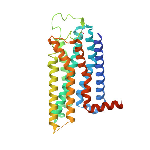
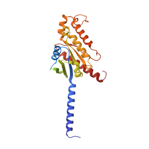
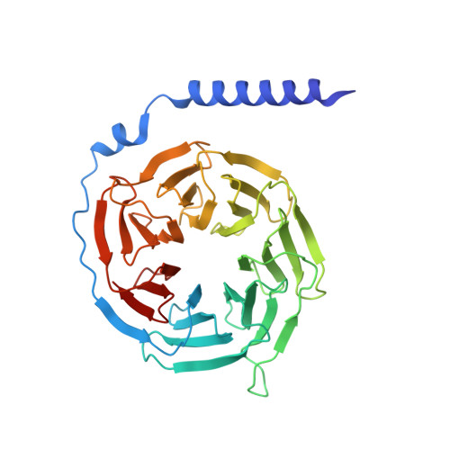
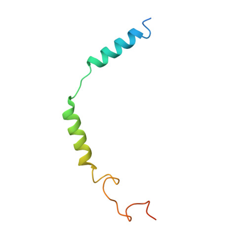
Cryo-EM structures of adenosine receptor A 3 AR bound to selective agonists.

Cai, H.Guo, S.Xu, Y.Sun, J.Li, J.Xia, Z.Jiang, Y.Xie, X.Xu, H.E.

(2024) Nat Commun 15: 3252-3252

  • DOI: https://doi.org/10.1038/s41467-024-47207-6
  • Primary Citation of Related Structures:  
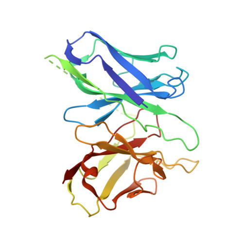
    8X16, 8X17

  • PubMed Abstract: 

    The adenosine A 3 receptor (A 3 AR), a key member of the G protein-coupled receptor family, is a promising therapeutic target for inflammatory and cancerous conditions. The selective A 3 AR agonists, CF101 and CF102, are clinically significant, yet their recognition mechanisms remained elusive. Here we report the cryogenic electron microscopy structures of the full-length human A 3 AR bound to CF101 and CF102 with heterotrimeric G i protein in complex at 3.3-3.2 Å resolution. These agonists reside in the orthosteric pocket, forming conserved interactions via their adenine moieties, while their 3-iodobenzyl groups exhibit distinct orientations. Functional assays reveal the critical role of extracellular loop 3 in A 3 AR's ligand selectivity and receptor activation. Key mutations, including His 3.37 , Ser 5.42 , and Ser 6.52 , in a unique sub-pocket of A 3 AR, significantly impact receptor activation. Comparative analysis with the inactive A 2A AR structure highlights a conserved receptor activation mechanism. Our findings provide comprehensive insights into the molecular recognition and signaling of A 3 AR, paving the way for designing subtype-selective adenosine receptor ligands.


  • Organizational Affiliation

    State Key Laboratory of Drug Research, Shanghai Institute of Materia Medica, Chinese Academy of Sciences, Shanghai, China. caihongmin@simm.ac.cn.


Macromolecules
Find similar proteins by:  (by identity cutoff)  |  3D Structure
Entity ID: 1
MoleculeChains Sequence LengthOrganismDetailsImage
Adenosine receptor A3A [auth R]318Homo sapiensMutation(s): 0 
Gene Names: ADORA3
Membrane Entity: Yes 
UniProt & NIH Common Fund Data Resources
Find proteins for P0DMS8 (Homo sapiens)
Explore P0DMS8 
Go to UniProtKB:  P0DMS8
PHAROS:  P0DMS8
GTEx:  ENSG00000282608 
Entity Groups  
Sequence Clusters30% Identity50% Identity70% Identity90% Identity95% Identity100% Identity
UniProt GroupP0DMS8
Sequence Annotations
Expand
  • Reference Sequence
Find similar proteins by:  (by identity cutoff)  |  3D Structure
Entity ID: 2
MoleculeChains Sequence LengthOrganismDetailsImage
Guanine nucleotide-binding protein G(i) subunit alpha-1B [auth A]354Bos taurusMutation(s): 2 
Gene Names: GNAI1
Membrane Entity: Yes 
UniProt
Find proteins for P63097 (Bos taurus)
Explore P63097 
Go to UniProtKB:  P63097
Entity Groups  
Sequence Clusters30% Identity50% Identity70% Identity90% Identity95% Identity100% Identity
UniProt GroupP63097
Sequence Annotations
Expand
  • Reference Sequence
Find similar proteins by:  (by identity cutoff)  |  3D Structure
Entity ID: 3
MoleculeChains Sequence LengthOrganismDetailsImage
Guanine nucleotide-binding protein G(I)/G(S)/G(T) subunit beta-1C [auth B]345Rattus norvegicusMutation(s): 0 
Gene Names: Gnb1
UniProt
Find proteins for P54311 (Rattus norvegicus)
Explore P54311 
Go to UniProtKB:  P54311
Entity Groups  
Sequence Clusters30% Identity50% Identity70% Identity90% Identity95% Identity100% Identity
UniProt GroupP54311
Sequence Annotations
Expand
  • Reference Sequence
Find similar proteins by:  (by identity cutoff)  |  3D Structure
Entity ID: 4
MoleculeChains Sequence LengthOrganismDetailsImage
Guanine nucleotide-binding protein G(I)/G(S)/G(O) subunit gamma-2D [auth C]71Bos taurusMutation(s): 0 
Gene Names: GNG2
Membrane Entity: Yes 
UniProt
Find proteins for P63212 (Bos taurus)
Explore P63212 
Go to UniProtKB:  P63212
Entity Groups  
Sequence Clusters30% Identity50% Identity70% Identity90% Identity95% Identity100% Identity
UniProt GroupP63212
Sequence Annotations
Expand
  • Reference Sequence
Find similar proteins by:  (by identity cutoff)  |  3D Structure
Entity ID: 5
MoleculeChains Sequence LengthOrganismDetailsImage
scFv16E [auth H]247Mus musculusMutation(s): 0 
Entity Groups  
Sequence Clusters30% Identity50% Identity70% Identity90% Identity95% Identity100% Identity
Sequence Annotations
Expand
  • Reference Sequence
Small Molecules
Ligands 1 Unique
IDChains Name / Formula / InChI Key2D Diagram3D Interactions
Q8L (Subject of Investigation/LOI)
Query on Q8L

Download Ideal Coordinates CCD File 
F [auth R]Piclidenoson
C18 H19 I N6 O4
HUJXGQILHAUCCV-MOROJQBDSA-N
Experimental Data & Validation

Experimental Data

  • Method: ELECTRON MICROSCOPY
  • Resolution: 3.29 Å
  • Aggregation State: PARTICLE 
  • Reconstruction Method: SINGLE PARTICLE 

Structure Validation

View Full Validation Report


Entry History & Funding Information

Deposition Data


Funding OrganizationLocationGrant Number
National Natural Science Foundation of China (NSFC)China81902085

Revision History  (Full details and data files)

  • Version 1.0: 2024-04-24
    Type: Initial release
  • Version 1.1: 2024-05-08
    Changes: Database references