The Conformational Change of the L3 Loop Affects the Structural Changes in the Substrate Binding Pocket Entrance of beta-Glucosidase.
Nam, K.H.(2023) Molecules 28
- PubMed: 38067537
- DOI: https://doi.org/10.3390/molecules28237807
- Primary Citation of Related Structures:
8WFT, 8WFU, 8WFV, 8WFW - PubMed Abstract:
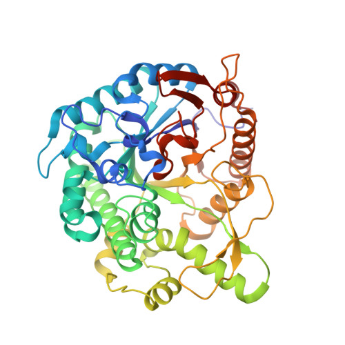
β-glucosidase (Bgl) hydrolyzes cellobiose to glucose, thereby releasing non-reducing terminal glucosyl residues. Bgl is an essential enzyme belonging to the biomass-degrading enzyme family, which plays a vital role in enzymatic saccharification during biofuel production. The four loops above the Bgl substrate-binding pocket undergo a conformational change upon substrate recognition. However, the structural dynamism of this loop and how it is conserved among Bgl family members remain unknown. Herein, to better understand the four loops above the substrate-binding pocket of Bgl, four Bgl crystal structures in Thermoanaerobacterium saccharolyticum (TsaBgl) were determined at 1.5-2.1 Å. The L1, L2, and L4 loops of TsaBgl showed a rigid conformation stabilized by their neighboring residues via hydrogen bonds and hydrophobic interactions. The TsaBgl L3 loop showed relatively high flexibility and two different N-terminal region conformations. The conformational change in the TsaBgl L3 loop induced a change in charge and shaped at the substrate-binding pocket entrance. The amino acid sequences and structures of the TsaBgl L1-4 loops were compared with other 45 Bgl proteins, and a diversity of the L2 and L3 loops was observed. Differences in amino acids and lengths of Bgls L2-L3 loop induced differences in the conformation and structure of the Bgls substrate-binding pocket entrance. These findings expand our knowledge on the molecular function of the loops in the Bgl enzyme family.
Organizational Affiliation:
College of General Education, Kookmin University, Seoul 02707, Republic of Korea.