8SMT

Crystal structure of antibody WRAIR-2134 in complex with SARS-CoV-2 receptor binding domain


Experimental Data Snapshot

  • Method: X-RAY DIFFRACTION
  • Resolution: 3.16 Å
  • R-Value Free: 0.249 
  • R-Value Work: 0.214 
  • R-Value Observed: 0.216 

wwPDB Validation   3D Report Full Report


Ligand Structure Quality Assessment 


This is version 1.2 of the entry. See complete history


Literature

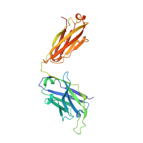
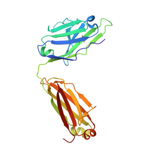
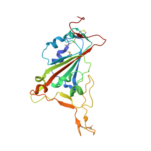
Antibody targeting of conserved sites of vulnerability on the SARS-CoV-2 spike receptor-binding domain.

Sankhala, R.S.Dussupt, V.Chen, W.H.Bai, H.Martinez, E.J.Jensen, J.L.Rees, P.A.Hajduczki, A.Chang, W.C.Choe, M.Yan, L.Sterling, S.L.Swafford, I.Kuklis, C.Soman, S.King, J.Corbitt, C.Zemil, M.Peterson, C.E.Mendez-Rivera, L.Townsley, S.M.Donofrio, G.C.Lal, K.G.Tran, U.Green, E.C.Smith, C.de Val, N.Laing, E.D.Broder, C.C.Currier, J.R.Gromowski, G.D.Wieczorek, L.Rolland, M.Paquin-Proulx, D.van Dyk, D.Britton, Z.Rajan, S.Loo, Y.M.McTamney, P.M.Esser, M.T.Polonis, V.R.Michael, N.L.Krebs, S.J.Modjarrad, K.Joyce, M.G.

(2024) Structure 32: 131

  • DOI: https://doi.org/10.1016/j.str.2023.11.015
  • Primary Citation of Related Structures:  
    7U8E, 8FAH, 8SGU, 8SMI, 8SMT

  • PubMed Abstract: 

    Given the continuous emergence of severe acute respiratory syndrome coronavirus 2 (SARS-CoV-2) variants of concern (VoCs), immunotherapeutics that target conserved epitopes on the spike (S) glycoprotein have therapeutic advantages. Here, we report the crystal structure of the SARS-CoV-2 S receptor-binding domain (RBD) at 1.95 Å and describe flexibility and distinct conformations of the angiotensin-converting enzyme 2 (ACE2)-binding site. We identify a set of SARS-CoV-2-reactive monoclonal antibodies (mAbs) with broad RBD cross-reactivity including SARS-CoV-2 Omicron subvariants, SARS-CoV-1, and other sarbecoviruses and determine the crystal structures of mAb-RBD complexes with Ab246 and CR3022 mAbs targeting the class IV site, WRAIR-2134, which binds the recently designated class V epitope, and WRAIR-2123, the class I ACE2-binding site. The broad reactivity of class IV and V mAbs to conserved regions of SARS-CoV-2 VoCs and other sarbecovirus provides a framework for long-term immunotherapeutic development strategies.


  • Organizational Affiliation

    Emerging Infectious Diseases Branch, Walter Reed Army Institute of Research, Silver Spring, MD, USA; Henry M. Jackson Foundation for the Advancement of Military Medicine, Bethesda, MD, USA.


Macromolecules
Find similar proteins by:  (by identity cutoff)  |  3D Structure
Entity ID: 1
MoleculeChains Sequence LengthOrganismDetailsImage
WRAIR-2134 Fab heavy chainA,
C [auth D],
E [auth H],
F [auth I]
231Homo sapiensMutation(s): 0 
Entity Groups  
Sequence Clusters30% Identity50% Identity70% Identity90% Identity95% Identity100% Identity
Sequence Annotations
Expand
  • Reference Sequence
Find similar proteins by:  (by identity cutoff)  |  3D Structure
Entity ID: 2
MoleculeChains Sequence LengthOrganismDetailsImage
WRAIR-2134 Fab light chainB,
D [auth F],
G [auth J],
H [auth L]
214Homo sapiensMutation(s): 0 
Entity Groups  
Sequence Clusters30% Identity50% Identity70% Identity90% Identity95% Identity100% Identity
Sequence Annotations
Expand
  • Reference Sequence
Find similar proteins by:  (by identity cutoff)  |  3D Structure
Entity ID: 3
MoleculeChains Sequence LengthOrganismDetailsImage
Spike protein S1I [auth C],
J [auth E],
K [auth G],
L [auth K]
205Severe acute respiratory syndrome coronavirus 2Mutation(s): 0 
Gene Names: S2
UniProt
Find proteins for P0DTC2 (Severe acute respiratory syndrome coronavirus 2)
Explore P0DTC2 
Go to UniProtKB:  P0DTC2
Entity Groups  
Sequence Clusters30% Identity50% Identity70% Identity90% Identity95% Identity100% Identity
UniProt GroupP0DTC2
Sequence Annotations
Expand
  • Reference Sequence
Oligosaccharides

Help

Entity ID: 4
MoleculeChains Length2D Diagram Glycosylation3D Interactions
beta-D-mannopyranose-(1-4)-beta-D-mannopyranose-(1-4)-2-acetamido-2-deoxy-beta-D-glucopyranose
M
3N-Glycosylation
Entity ID: 5
MoleculeChains Length2D Diagram Glycosylation3D Interactions
beta-D-mannopyranose-(1-4)-2-acetamido-2-deoxy-beta-D-glucopyranose-(1-4)-2-acetamido-2-deoxy-beta-D-glucopyranose
N
3N-Glycosylation
Glycosylation Resources
GlyTouCan:  G15407YE
GlyCosmos:  G15407YE
GlyGen:  G15407YE
Small Molecules
Ligands 1 Unique
IDChains Name / Formula / InChI Key2D Diagram3D Interactions
NAG
Query on NAG

Download Ideal Coordinates CCD File 
O [auth C],
P [auth E]
2-acetamido-2-deoxy-beta-D-glucopyranose
C8 H15 N O6
OVRNDRQMDRJTHS-FMDGEEDCSA-N
Experimental Data & Validation

Experimental Data

  • Method: X-RAY DIFFRACTION
  • Resolution: 3.16 Å
  • R-Value Free: 0.249 
  • R-Value Work: 0.214 
  • R-Value Observed: 0.216 
  • Space Group: P 21 21 21
Unit Cell:
Length ( Å )Angle ( ˚ )
a = 143.19α = 90
b = 154.43β = 90
c = 165.25γ = 90
Software Package:
Software NamePurpose
PHENIXrefinement
HKL-2000data reduction
HKL-2000data scaling
PHASERphasing

Structure Validation

View Full Validation Report


Ligand Structure Quality Assessment 


Entry History & Funding Information

Deposition Data


Funding OrganizationLocationGrant Number
Department of Defense (DOD, United States)United StatesW81XWH-18-2-0040

Revision History  (Full details and data files)

  • Version 1.0: 2023-06-28
    Type: Initial release
  • Version 1.1: 2024-01-10
    Changes: Data collection, Database references
  • Version 1.2: 2024-02-14
    Changes: Database references