8SLV

Structure of a salivary alpha-glucosidase from the mosquito vector Aedes aegypti.


Experimental Data Snapshot

  • Method: X-RAY DIFFRACTION
  • Resolution: 2.32 Å
  • R-Value Free: 0.188 
  • R-Value Work: 0.146 
  • R-Value Observed: 0.149 

wwPDB Validation   3D Report Full Report


Ligand Structure Quality Assessment 


This is version 1.0 of the entry. See complete history


Literature

Structural and functional comparisons of salivary alpha-glucosidases from the mosquito vectors Aedes aegypti, Anopheles gambiae, and Culex quinquefasciatus.

Williams, A.E.Gittis, A.G.Botello, K.Cruz, P.Martin-Martin, I.Valenzuela Leon, P.C.Sumner, B.Bonilla, B.Calvo, E.

(2024) Insect Biochem Mol Biol 167: 104097-104097

  • DOI: https://doi.org/10.1016/j.ibmb.2024.104097
  • Primary Citation of Related Structures:  
    8SLV

  • PubMed Abstract: 

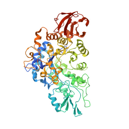
    Mosquito vectors of medical importance both blood and sugar feed, and their saliva contains bioactive molecules that aid in both processes. Although it has been shown that the salivary glands of several mosquito species exhibit α-glucosidase activities, the specific enzymes responsible for sugar digestion remain understudied. We therefore expressed and purified three recombinant salivary α-glucosidases from the mosquito vectors Aedes aegypti, Anopheles gambiae, and Culex quinquefasciatus and compared their functions and structures. We found that all three enzymes were expressed in the salivary glands of their respective vectors and were secreted into the saliva. The proteins, as well as mosquito salivary gland extracts, exhibited α-glucosidase activity, and the recombinant enzymes displayed preference for sucrose compared to p-nitrophenyl-α-D-glucopyranoside. Finally, we solved the crystal structure of the Ae. aegypti α-glucosidase bound to two calcium ions at a 2.3 Ångstrom resolution. Molecular docking suggested that the Ae. aegypti α-glucosidase preferred di- or polysaccharides compared to monosaccharides, consistent with enzymatic activity assays. Comparing structural models between the three species revealed a high degree of similarity, suggesting similar functional properties. We conclude that the α-glucosidases studied herein are important enzymes for sugar digestion in three mosquito species.


  • Organizational Affiliation

    Laboratory of Malaria and Vector Research, National Institute of Allergy and Infectious Diseases, National Institutes of Health, Rockville, MD, 20852, USA.


Macromolecules
Find similar proteins by:  (by identity cutoff)  |  3D Structure
Entity ID: 1
MoleculeChains Sequence LengthOrganismDetailsImage
Salivary alpha-glucosidase579Aedes aegyptiMutation(s): 0 
Gene Names: MAL1MAL IAAEL000392
EC: 3.2.1.20
UniProt
Find proteins for P13080 (Aedes aegypti)
Explore P13080 
Go to UniProtKB:  P13080
Entity Groups  
Sequence Clusters30% Identity50% Identity70% Identity90% Identity95% Identity100% Identity
UniProt GroupP13080
Sequence Annotations
Expand
  • Reference Sequence
Small Molecules
Ligands 8 Unique
IDChains Name / Formula / InChI Key2D Diagram3D Interactions
NAG
Query on NAG

Download Ideal Coordinates CCD File 
B [auth A]2-acetamido-2-deoxy-beta-D-glucopyranose
C8 H15 N O6
OVRNDRQMDRJTHS-FMDGEEDCSA-N
TRS
Query on TRS

Download Ideal Coordinates CCD File 
I [auth A]2-AMINO-2-HYDROXYMETHYL-PROPANE-1,3-DIOL
C4 H12 N O3
LENZDBCJOHFCAS-UHFFFAOYSA-O
CYS
Query on CYS

Download Ideal Coordinates CCD File 
C [auth A]CYSTEINE
C3 H7 N O2 S
XUJNEKJLAYXESH-REOHCLBHSA-N
GOL
Query on GOL

Download Ideal Coordinates CCD File 
D [auth A],
E [auth A],
F [auth A],
G [auth A],
H [auth A]
GLYCEROL
C3 H8 O3
PEDCQBHIVMGVHV-UHFFFAOYSA-N
PDO
Query on PDO

Download Ideal Coordinates CCD File 
K [auth A]1,3-PROPANDIOL
C3 H8 O2
YPFDHNVEDLHUCE-UHFFFAOYSA-N
IPA
Query on IPA

Download Ideal Coordinates CCD File 
J [auth A]ISOPROPYL ALCOHOL
C3 H8 O
KFZMGEQAYNKOFK-UHFFFAOYSA-N
CA
Query on CA

Download Ideal Coordinates CCD File 
L [auth A],
M [auth A]
CALCIUM ION
Ca
BHPQYMZQTOCNFJ-UHFFFAOYSA-N
CL
Query on CL

Download Ideal Coordinates CCD File 
N [auth A],
O [auth A],
P [auth A],
Q [auth A]
CHLORIDE ION
Cl
VEXZGXHMUGYJMC-UHFFFAOYSA-M
Experimental Data & Validation

Experimental Data

  • Method: X-RAY DIFFRACTION
  • Resolution: 2.32 Å
  • R-Value Free: 0.188 
  • R-Value Work: 0.146 
  • R-Value Observed: 0.149 
  • Space Group: P 1
Unit Cell:
Length ( Å )Angle ( ˚ )
a = 48.76α = 101.816
b = 51.03β = 94.658
c = 70.75γ = 95.764
Software Package:
Software NamePurpose
PHENIXrefinement
PHENIXrefinement
XDSdata reduction
XSCALEdata scaling
PHENIXphasing

Structure Validation

View Full Validation Report


Ligand Structure Quality Assessment 


Entry History & Funding Information

Deposition Data


Funding OrganizationLocationGrant Number
National Institutes of Health/National Institute Of Allergy and Infectious Diseases (NIH/NIAID)United States--

Revision History  (Full details and data files)

  • Version 1.0: 2024-03-20
    Type: Initial release