8RHI

Lytic Transglycosylase MltD of Pseudomonas aeruginosa in a ternary complex bound to Bulgecin A and chito-tetraose


Experimental Data Snapshot

  • Method: X-RAY DIFFRACTION
  • Resolution: 1.98 Å
  • R-Value Free: 0.215 
  • R-Value Work: 0.190 

wwPDB Validation   3D Report Full Report


Ligand Structure Quality Assessment 


This is version 1.1 of the entry. See complete history


Literature

Structural characterization of lytic transglycosylase MltD of Pseudomonas aeruginosa, a target for the natural product bulgecin A.

Miguel-Ruano, V.Feltzer, R.Batuecas, M.T.Ramachandran, B.El-Araby, A.M.Avila-Cobian, L.F.De Benedetti, S.Mobashery, S.Hermoso, J.A.

(2024) Int J Biol Macromol 267: 131420-131420

  • DOI: https://doi.org/10.1016/j.ijbiomac.2024.131420
  • Primary Citation of Related Structures:  
    8RHE, 8RHF, 8RHI

  • PubMed Abstract: 

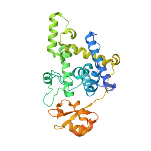
    Natural product bulgecin A potentiates the activity of β-lactam antibiotics by inhibition of three lytic transglycosylases in Pseudomonas aeruginosa, of which MltD is one. MltD exhibits both endolytic and exolytic reactions in the turnover of the cell-wall peptidoglycan and tolerates the presence or absence of stem peptides in its substrates. The present study reveals structural features of the multimodular MltD, presenting a catalytic module and four cell-wall-binding LysM modules that account for these attributes. Three X-ray structures are reported herein for MltD that disclose one unpredicted LysM module tightly attached to the catalytic domain, whereas the other LysM modules are mobile, and connected to the catalytic domain through long flexible linkers. The formation of crystals depended on the presence of bulgecin A. The expansive active-site cleft is highlighted by the insertion of a helical region, a hallmark of the family 1D of lytic transglycosylases, which was mapped out in a ternary complex of MltD:bulgecinA:chitotetraose, revealing at the minimum the presence of eight subsites (from -4 to +4, with the seat of reaction at subsites -1 and + 1) for binding of sugars of the substrate for the endolytic reaction. The mechanism of the exolytic reaction is revealed in one of the structures, showing how the substrate's terminal anhydro-NAM moiety could be sequestered at subsite +2. Our results provide the structural insight for both the endolytic and exolytic activities of MltD during cell-wall-turnover events.


  • Organizational Affiliation

    Department of Crystallography and Structural Biology, Instituto de Química-Física "Blas Cabrera", Consejo Superior de Investigaciones Científicas, Madrid, Spain.


Macromolecules
Find similar proteins by:  (by identity cutoff)  |  3D Structure
Entity ID: 1
MoleculeChains Sequence LengthOrganismDetailsImage
LysM peptidoglycan-binding domain-containing protein
A, B
338Pseudomonas aeruginosaMutation(s): 0 
Gene Names: mltDCAZ10_25765CGU42_31890GNQ48_00460GUL26_19730IPC1295_05870PAERUG_P19_London_7_VIM_2_05_10_06006
EC: 4.2.2
UniProt
Find proteins for A0A0C7CWY9 (Pseudomonas aeruginosa)
Explore A0A0C7CWY9 
Go to UniProtKB:  A0A0C7CWY9
Entity Groups  
Sequence Clusters30% Identity50% Identity70% Identity90% Identity95% Identity100% Identity
UniProt GroupA0A0C7CWY9
Sequence Annotations
Expand
  • Reference Sequence
Oligosaccharides

Help

Entity ID: 2
MoleculeChains Length2D Diagram Glycosylation3D Interactions
2-acetamido-2-deoxy-beta-D-glucopyranose-(1-4)-2-acetamido-2-deoxy-beta-D-glucopyranoseC [auth D]2N/A
Glycosylation Resources
GlyTouCan:  G42666HT
GlyCosmos:  G42666HT
GlyGen:  G42666HT
Entity ID: 3
MoleculeChains Length2D Diagram Glycosylation3D Interactions
2-acetamido-2-deoxy-beta-D-glucopyranose-(1-4)-2-acetamido-2-deoxy-beta-D-glucopyranose-(1-4)-2-acetamido-2-deoxy-beta-D-glucopyranose-(1-4)-2-acetamido-2-deoxy-beta-D-glucopyranoseD [auth E]4N/A
Glycosylation Resources
GlyTouCan:  G01361SX
GlyCosmos:  G01361SX
GlyGen:  G01361SX
Small Molecules
Ligands 2 Unique
IDChains Name / Formula / InChI Key2D Diagram3D Interactions
BLG (Subject of Investigation/LOI)
Query on BLG

Download Ideal Coordinates CCD File 
E [auth A],
L [auth B]
4-O-(4-O-SULFONYL-N-ACETYLGLUCOSAMININYL)-5-METHYLHYDROXY-L-PROLINE-TAURINE
C16 H30 N3 O14 S2
RPNZWZDLNYCCIG-HMMVDTEZSA-O
ZN
Query on ZN

Download Ideal Coordinates CCD File 
F [auth A]
G [auth A]
H [auth A]
I [auth A]
J [auth A]
F [auth A],
G [auth A],
H [auth A],
I [auth A],
J [auth A],
K [auth A],
M [auth B],
N [auth B],
O [auth B]
ZINC ION
Zn
PTFCDOFLOPIGGS-UHFFFAOYSA-N
Experimental Data & Validation

Experimental Data

  • Method: X-RAY DIFFRACTION
  • Resolution: 1.98 Å
  • R-Value Free: 0.215 
  • R-Value Work: 0.190 
  • Space Group: P 21 21 21
Unit Cell:
Length ( Å )Angle ( ˚ )
a = 45.248α = 90
b = 117.036β = 90
c = 120.146γ = 90
Software Package:
Software NamePurpose
REFMACrefinement
XDSdata reduction
Aimlessdata scaling
PHASERphasing

Structure Validation

View Full Validation Report


Ligand Structure Quality Assessment 


Entry History & Funding Information

Deposition Data


Funding OrganizationLocationGrant Number
Spanish Ministry of Science, Innovation, and UniversitiesSpainBFU2017-90030-P

Revision History  (Full details and data files)

  • Version 1.0: 2024-04-17
    Type: Initial release
  • Version 1.1: 2024-04-24
    Changes: Database references