8RF0

WT-CGS sample in nanodisc


Experimental Data Snapshot

  • Method: ELECTRON MICROSCOPY
  • Resolution: 3.40 Å
  • Aggregation State: PARTICLE 
  • Reconstruction Method: SINGLE PARTICLE 

wwPDB Validation   3D Report Full Report


This is version 1.0 of the entry. See complete history


Literature

Structure-function analysis of the cyclic beta-1,2-glucan synthase from Agrobacterium tumefaciens.

Sedzicki, J.Ni, D.Lehmann, F.Stahlberg, H.Dehio, C.

(2024) Nat Commun 15: 1844

  • DOI: https://doi.org/10.1038/s41467-024-45415-8
  • Primary Citation of Related Structures:  
    8RF0, 8RF9, 8RFE, 8RFG

  • PubMed Abstract: 

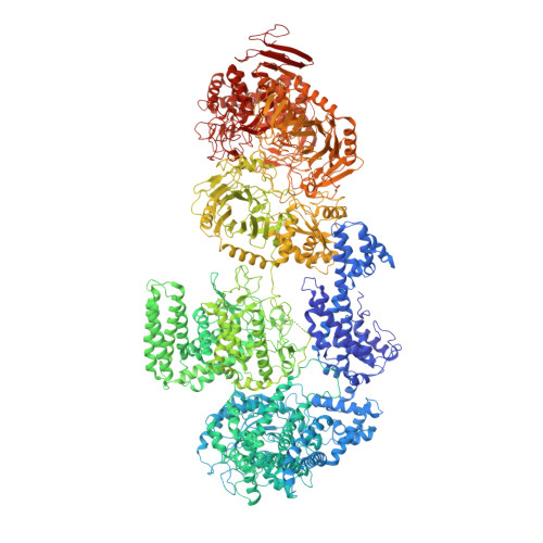
    The synthesis of complex sugars is a key aspect of microbial biology. Cyclic β-1,2-glucan (CβG) is a circular polysaccharide critical for host interactions of many bacteria, including major pathogens of humans (Brucella) and plants (Agrobacterium). CβG is produced by the cyclic glucan synthase (Cgs), a multi-domain membrane protein. So far, its structure as well as the mechanism underlining the synthesis have not been clarified. Here we use cryo-electron microscopy (cryo-EM) and functional approaches to study Cgs from A. tumefaciens. We determine the structure of this complex protein machinery and clarify key aspects of CβG synthesis, revealing a distinct mechanism that uses a tyrosine-linked oligosaccharide intermediate in cycles of polymerization and processing of the glucan chain. Our research opens possibilities for combating pathogens that rely on polysaccharide virulence factors and may lead to synthetic biology approaches for producing complex cyclic sugars.


  • Organizational Affiliation

    Biozentrum, University of Basel, Basel, CH-4056, Switzerland.


Macromolecules
Find similar proteins by:  (by identity cutoff)  |  3D Structure
Entity ID: 1
MoleculeChains Sequence LengthOrganismDetailsImage
Cyclic beta-(1,2)-glucan synthase NdvB2,818Agrobacterium tumefaciensMutation(s): 0 
Gene Names: ndvBAGTUEHA105_LOCUS2378
Membrane Entity: Yes 
UniProt
Find proteins for A0A6V6ZZ23 (Rhizobium radiobacter)
Explore A0A6V6ZZ23 
Go to UniProtKB:  A0A6V6ZZ23
Entity Groups  
Sequence Clusters30% Identity50% Identity70% Identity90% Identity95% Identity100% Identity
UniProt GroupA0A6V6ZZ23
Sequence Annotations
Expand
  • Reference Sequence
Oligosaccharides

Help

Entity ID: 2
MoleculeChains Length2D Diagram Glycosylation3D Interactions
beta-D-glucopyranose-(1-2)-beta-D-glucopyranose-(1-2)-beta-D-glucopyranose-(1-2)-beta-D-glucopyranose-(1-2)-beta-D-glucopyranose-(1-2)-beta-D-glucopyranose-(1-2)-beta-D-glucopyranose-(1-2)-beta-D-glucopyranose-(1-2)-beta-D-glucopyranose
B
9N/A
Entity ID: 3
MoleculeChains Length2D Diagram Glycosylation3D Interactions
beta-D-glucopyranose-(1-2)-beta-D-glucopyranose-(1-2)-beta-D-glucopyranose-(1-2)-beta-D-glucopyranose-(1-2)-beta-D-glucopyranose-(1-3)-beta-D-glucopyranose-(1-2)-beta-D-glucopyranose-(1-2)-beta-D-glucopyranose-(1-3)-beta-D-glucopyranose
C
9N/A
Small Molecules
Ligands 1 Unique
IDChains Name / Formula / InChI Key2D Diagram3D Interactions
UPG
Query on UPG

Download Ideal Coordinates CCD File 
D [auth A]URIDINE-5'-DIPHOSPHATE-GLUCOSE
C15 H24 N2 O17 P2
HSCJRCZFDFQWRP-JZMIEXBBSA-N
Experimental Data & Validation

Experimental Data

  • Method: ELECTRON MICROSCOPY
  • Resolution: 3.40 Å
  • Aggregation State: PARTICLE 
  • Reconstruction Method: SINGLE PARTICLE 

Structure Validation

View Full Validation Report


Entry History & Funding Information

Deposition Data


Funding OrganizationLocationGrant Number
Swiss National Science FoundationSwitzerland310030B_201273
Swiss National Science FoundationSwitzerlandNCCR AntiResist grant 180541
Swiss National Science FoundationSwitzerlandCRSII5_177195
Swiss National Science FoundationSwitzerlandSino-Swiss Science and Technology Cooperation SSSTC grant

Revision History  (Full details and data files)

  • Version 1.0: 2024-03-20
    Type: Initial release